
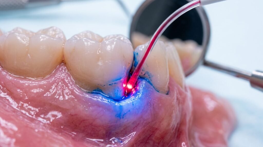
Antimicrobial resistance among subgingival periodontal pathogens poses an escalating threat to conventional non-surgical and adjunctive pharmacotherapies. Over decades of clinical practice, systemic administration of amoxicillin, metronidazole, and tetracyclines has generated resistant strains of Porphyromonas gingivalis, Tannerella forsythia, and Aggregatibacter actinomycetemcomitans within subgingival biofilm ecosystems. Furthermore, systemic antimicrobials frequently cause gastrointestinal dysbiosis, allergic hypersensitivity, and insufficient active drug concentrations within avascular, crevicular fluid environments. Antimicrobial photodynamic therapy provides a localized, non-pharmacologic biocidal modality capable of eradicating recalcitrant anaerobic pathogens instantaneously. Utilizing non-toxic photosensitizing dyes, such as phenothiazine chloride, methylene blue, or indocyanine green, activated by specific low-power visible or near-infrared laser light, this photochemical reaction generates lethal quantities of singlet oxygen and hydroxyl free radicals. These reactive oxygen species disrupt microbial cell membranes and intracellular organelles through unselective oxidative stress, destroying complex polymicrobial biofilms without inducing bacterial resistance.
Photochemical Principles and Singlet Oxygen Cytotoxicity
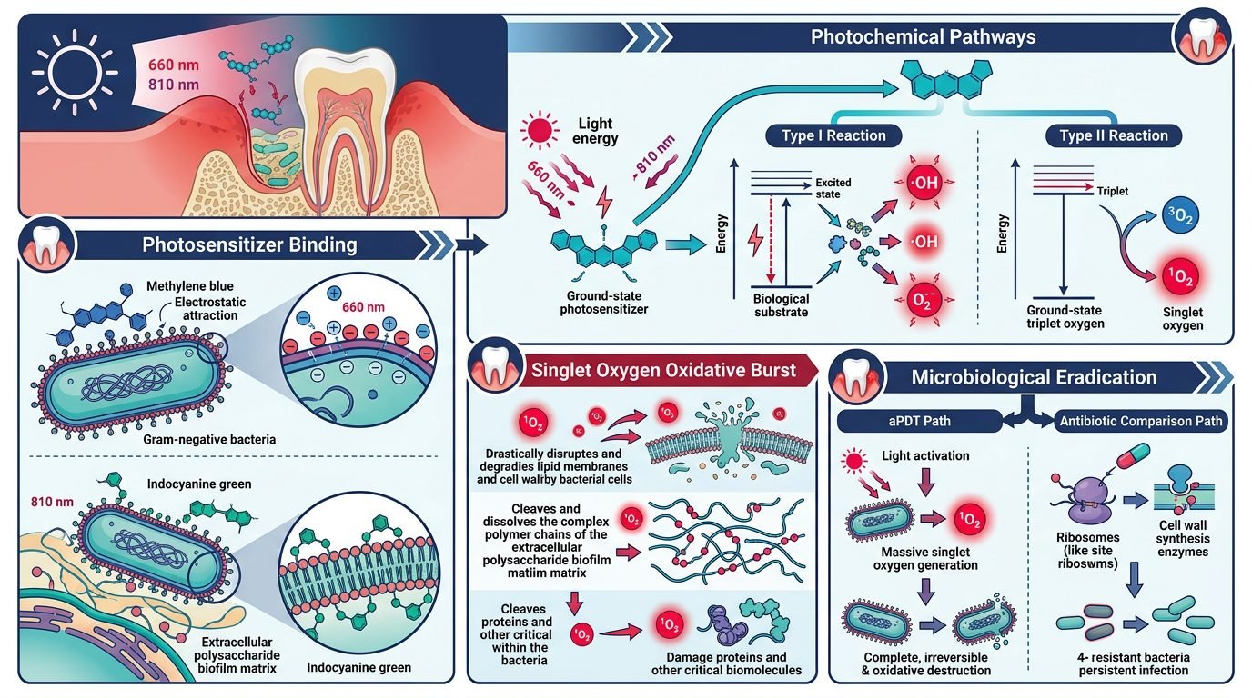
The biophysical foundation of antimicrobial photodynamic therapy rests upon three non-toxic components: light of a matching optical resonance wavelength, a chemical photosensitizer, and dissolved ambient ground-state molecular oxygen. When a photosensitizer molecule absorbs a photon matching its specific electronic absorption band, it transitions from a low-energy ground singlet state to an excited singlet state. Through intersystem crossing, the excited molecule undergoes spin inversion, entering an excited triplet state with a substantially longer microsecond lifespan.
In the triplet state, the photosensitizer interacts with neighboring substrates via two competing photochemical pathways. In the type one reaction pathway, electron or hydrogen transfer occurs directly with adjacent microbial membrane lipids and proteins, producing toxic intermediate free radicals such as superoxide anions and hydroxyl radicals. In the type two reaction pathway, energy transfers directly from the triplet photosensitizer to surrounding ground-state triplet oxygen molecules. This spin-permitted energy transfer converts non-toxic ground-state oxygen into highly reactive singlet molecular oxygen. Singlet oxygen possesses an extraordinarily short biological half-life of less than forty nanoseconds and an active diffusion radius of fewer than twenty nanometers, restricting severe oxidative destruction strictly to the immediate microscopic vicinity of the sensitized bacterial membrane.
Photosensitizer Characteristics: Methylene Blue versus Indocyanine Green
The clinical efficacy of periodontal photo-inactivation depends heavily upon the chemical architecture and optical absorption properties of the selected photosensitizer dye. Methylene blue and toluidine blue O belong to the phenothiazinium class, carrying a permanent positive cationic charge. Because the outer cell envelopes of gram-negative periodontopathogens possess net negative electrostatic charges conferred by lipopolysaccharide phosphate groups, cationic phenothiazines bind avidly to outer membranes through spontaneous electrostatic attraction. Methylene blue exhibits peak electronic absorption between 660 and 670 nanometers, matching widely accessible visible red diode lasers.
In contrast, indocyanine green is a tricarbocyanine fluorophore with an optical absorption peak situated in the near-infrared spectrum between 800 and 810 nanometers. Indocyanine green provides exceptional tissue penetration depth, allowing therapeutic near-infrared photons to penetrate dense fibrous pocket walls and deep root concavities where red light experiences higher scattering. Additionally, indocyanine green generates both photodynamic oxidative cytotoxicity and localized photothermal lysis when excited by 810 nm diode radiation. Nanoparticle carriers and emulsion formulations enhance photosensitizer stability, preventing premature aqueous dye dimerization and ensuring maximal monomeric triplet state generation.
Biofilm Penetration and Eradication of Resistant Pathogens
Periodontopathogenic bacteria residing within established subgingival biofilms exhibit up to one thousand times higher resistance to conventional chemical antiseptics and systemic antibiotics compared to planktonic phenotypes. This profound protection stems from the dense extracellular polymeric substance matrix, composed of polysaccharides, extracellular DNA, and structural proteins that acts as a physical diffusion barrier. Traditional chlorhexidine irrigants and systemic tetracyclines fail to penetrate this viscous hydrogel shield effectively.
Antimicrobial photodynamic therapy overcomes biofilm shielding through non-specific, high-energy oxidative attack. When low-viscosity photosensitizer solutions are delivered into the pocket, capillary action draws the dye molecules into microscopic channels traversing the biofilm matrix. Subsequent laser activation produces an immediate explosion of singlet oxygen that cleaves extracellular polysaccharides, uncouples bacterial membrane lipopolysaccharides, and cross-links outer membrane porin proteins. Because singlet oxygen oxidizes multiple biochemical targets simultaneously, bacteria cannot develop genetic mutations or express efflux pumps to evade cytotoxicity. Laboratory testing confirms that repeated cycles of photodynamic exposure induce zero reduction in microbial susceptibility.
Clinical Protocols for Pocket Irrigation and Laser Irradiation
Predictable clinical success requires rigorous execution of a standardized subgingival application protocol following thorough mechanical scaling and root planing. Ultrasonic and hand instrumentation must first disrupt macroscopic calculus deposits and reduce dense biofilm volume. The subgingival pocket is then thoroughly flushed with sterile saline and gently dried with compressed air to prevent excess fluid from diluting the photosensitizer.
The photosensitizer dye is introduced into the bottom of the periodontal pocket utilizing a blunt, side-venting endodontic needle. An incubation period of sixty seconds allows the cationic dye to disperse through the remaining subgingival biofilm and bind electrostatically to bacterial envelopes. Following incubation, a flexible optical fiber handpiece (typically 200 to 300 micrometers in diameter) is inserted to the base of the pocket. The clinician irradiates the pocket in a continuous circumferential sweeping motion from apical to coronal depths. Power settings are maintained between 100 and 150 milliwatts, delivering an energy density of approximately 4 to 6 Joules per square centimeter per tooth site. Thorough saline post-lavage removes residual dye and necrotic debris.
Clinical Outcomes and Systemic Benefits
Numerous randomized controlled clinical trials and meta-analyses confirm that adjunctive antimicrobial photodynamic therapy produces significant clinical attachment gains and marked reductions in periodontal probing depths compared to mechanical scaling alone. Sites treated with photodynamic therapy display accelerated gingival bleeding index reductions and substantial suppression of key anaerobic pathogens up to six months post-intervention. Furthermore, the localized photochemical mechanism eliminates the risk of systemic side effects, such as opportunistic fungal overgrowth, hepatorenal strain, or drug-drug interactions common with systemic antibiotics.
For medically compromised patients, photodynamic therapy provides a safe, targeted option. The absence of systemic absorption shields host metabolism while neutralizing periodontal pathogens locally.
Significance in Modern Periodontal Practice
Antimicrobial photodynamic therapy represents an established biophysical milestone in non-antibiotic periodontal therapy. By substituting non-selective singlet oxygen cytotoxicity for systemic antimicrobial regimens, clinicians circumvent bacterial resistance mechanisms entirely. Precise subgingival light delivery, combined with biocompatible photosensitizers, preserves periodontal tissue architecture while achieving predictable microbiological suppression across deep periodontal defects.
References
- Sgolastra, F., et al. (2013). Photodynamic therapy in the treatment of chronic periodontitis: a systematic review and meta-analysis. Lasers in Medical Science, 28(2), 669-680.
- Meimandi, M., et al. (2017). The effect of photodynamic therapy in the treatment of chronic periodontitis: a review of literature. Journal of Lasers in Medical Sciences, 8(Suppl 1), S7-S11.
- Takasaki, A. A., et al. (2009). Application of antimicrobial photodynamic therapy in periodontal and peri-implant diseases. Periodontology 2000, 51(1), 109-140.
- Hamblin, M. R., & Hasan, T. (2004). Photodynamic therapy: a new antimicrobial approach to infectious disease? Photochemical & Photobiological Sciences, 3(5), 436-450.
- Andersen, R., et al. (2007). Adjunctive approach to conventional scaling and root planing using photodynamic therapy: a review. Journal of Clinical Dentistry, 18(2), 47-51.
Leave a Reply