
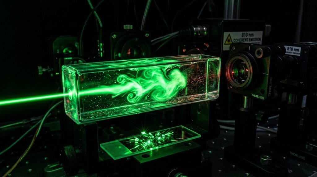
Antimicrobial photodynamic therapy (aPDT) utilizing indocyanine green (ICG) activated by near-infrared 810 nm diode lasers represents an advanced photochemical debridement adjunct for managing refractory peri-implantitis. Micro-rough, moderately sandblasted and acid-etched (SLA) titanium implant surfaces provide biocompatible topography for rapid osseointegration, but create impenetrable micro-niches for anaerobic biofilms once bone loss exposes implant threads to the oral environment. Mechanical curettes, titanium brushes, and ultrasonic scalers cannot conform to complex micro-threaded geometries without generating metal micro-particulates or leaving bacterial bio-burdens sequestered within deep thread valleys. Systemic antibiotics demonstrate declining efficacy due to bacterial beta-lactamase production and impaired subgingival vascular access around diseased implants. Traditional aPDT protocols relying on phenothiazine dyes, such as methylene blue or toluidine blue O activated by red light (660 nm), face severe biophysical hurdles, including limited optical tissue penetration and rapid photosensitizer degradation. Transitioning to near-infrared photosensitizers, specifically indocyanine green, significantly modifies photochemical penetration, reactive oxygen generation, and subgingival biofilm disruption.
The photochemical and photophysical distinctions between indocyanine green and phenothiazine dyes dictate clinical performance in deep peri-implant pockets. Methylene blue operates predominantly through Type II photochemical reactions, wherein photon absorption excites the dye into a triplet state that transfers energy directly to molecular ground-state oxygen, generating highly cytotoxic singlet oxygen (1O2). While singlet oxygen effectively lyses outer membranes of periodontal pathogens, methylene blue requires red excitation light around 660 nm. Blood, hemoglobin, and inflamed granulation tissue absorb 660 nm photons avidly, creating an optical extinction curtain that restricts therapeutic photon fluence to the uppermost one to two millimeters of bleeding peri-implant crevices. In refractory peri-implantitis cases featuring probing depths of six to ten millimeters and active bleeding, red light fails to penetrate down to the apical defect base.
Indocyanine green, an amphiphilic tricarbocyanine dye approved for intravenous vascular angiography, exhibits absorption peaks in the near-infrared spectrum between 800 nm and 820 nm. Near-infrared photons coincide with the biological optical window where tissue chromophore absorption is minimal, allowing photon penetration depths two to three times greater than red wavelengths through bleeding tissue. Furthermore, ICG functions through a dual photochemical and photothermal pathway. In addition to producing Type I (hydroxyl and superoxide radicals) and Type II reactive oxygen species, light absorption by ICG generates localized, micro-scale photothermal elevations within the photosensitizer film coating bacterial membranes. This localized photothermal spike destabilizes bacterial cell envelopes and denatures extracellular polymeric substances without conducting destructive heat to surrounding alveolar bone.
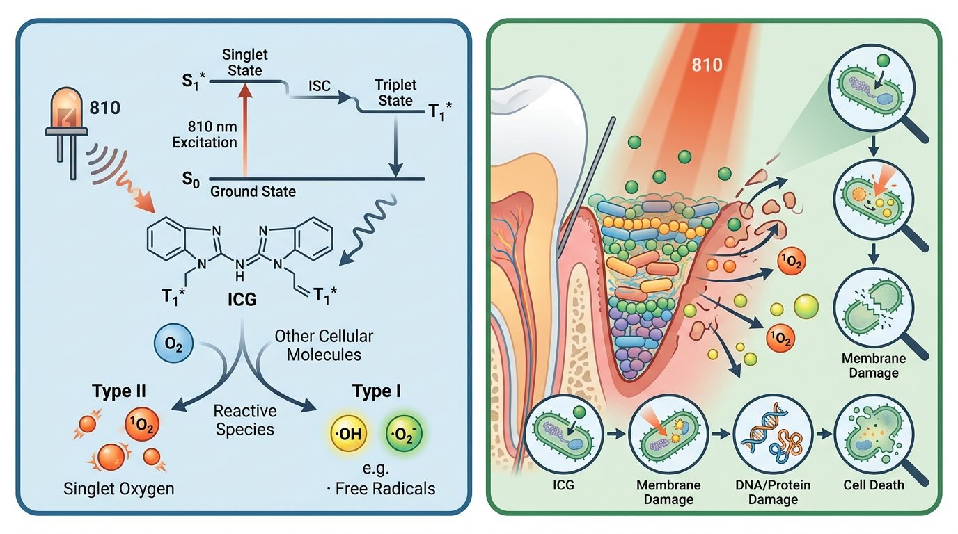
Efficacy against keystone peri-implant pathogens defines clinical utility. In vitro and clinical microbiologic investigations demonstrate that ICG-mediated aPDT achieves profound log-reductions in Porphyromonas gingivalis, Tannerella forsythia, Treponema denticola, and resistant Prevotella intermedia strains. Furthermore, ICG demonstrates high affinity for Candida albicans and Staphylococcus aureus, opportunistic pathogens frequently isolated from failing implant surfaces that show natural resistance to conventional periodontal therapies. The non-specific oxidative attack generated by aPDT damages bacterial cell membranes, disrupts essential transport proteins, and induces fatal DNA cleavage without risk of microbial resistance development, presenting a decisive biological advantage over repeated courses of systemic amoxicillin or metronidazole.
Formulation chemistry and pre-irradiation incubation parameters demand strict adherence. Unconjugated clinical-grade ICG powder must be freshly reconstituted with sterile aqueous diluent immediately prior to subgingival application, as aqueous ICG solutions undergo rapid hydrolytic degradation and photodegradation within hours of reconstitution. Concentrations between 0.5 mg/mL and 1.0 mg/mL (0.05 to 0.1 percent) provide an optimal balance between optical absorption and self-quenching phenomena. Excessively concentrated dye solutions create optical shielding, where superficial dye molecules absorb all incident photons and prevent radiation from reaching deeper bacterial layers. The subgingival incubation time must span sixty to ninety seconds, permitting the dye to coat contaminated implant threads, diffuse into subgingival calculus micro-crevices, and permeate the extracellular polysaccharide matrix before light activation begins.
Delivery kinetics and laser dosimetric parameters determine clinical success. Following thorough mechanical debridement with non-abrasive polyetheretherketone (PEEK) curettes or glycine powder air-polishing, the pocket is rinsed with sterile saline to clear loose debris and unseated blood clots. Freshly reconstituted ICG is delivered into the peri-implant pocket using a blunt-ended side-venting irrigation cannula until the pocket is gently backfilled. Activation requires an 810 nm diode laser delivered through a flexible optical fiber (three hundred to four hundred micrometers diameter) equipped with a diffusing tip. The fiber is introduced to within one millimeter of the pocket base and moved continuously in slow, circumferential sweeping motions around the implant circumference at a speed of approximately one to two millimeters per second. Power settings must remain strictly conservative: continuous wave output between 0.1 and 0.3 watts, or pulsed mode delivering average power of 0.2 watts, provides an irradiance that avoids thermal accumulation along the titanium fixture. The total irradiation time must average thirty to forty-five seconds per implant aspect (mesial, distal, buccal, lingual), totaling two to three minutes per affected fixture.
Longitudinal clinical trials comparing ICG-assisted aPDT with mechanical debridement alone confirm significant clinical improvements in clinical attachment levels, probing pocket depth reductions of 1.8 to 2.4 millimeters, and marked reductions in bleeding on probing (BOP) at six-month recalls. However, clinicians must recognize the biological limits of non-surgical decontamination. While ICG aPDT effectively suppresses microbial virulence and resolves mucosal inflammation, it does not regenerate lost circumferential or intrabony alveolar architecture. Where deep crater-like osseous defects persist, surgical access, mechanical implantoplasty, or regenerative bone grafting with barrier membranes remains necessary to reconstruct supporting structures.
Preservation of titanium surface integrity represents another compelling justification for selecting photochemical therapy over aggressive mechanical or ablative modalities. Conventional ultrasonic metal tips and high-speed rotary burs gouge the micro-machined titanium collar and strip the passivating titanium dioxide (TiO2) protective layer, releasing microscopic titanium particulates that incite local macrophage activation and accelerate osteoclast-mediated bone breakdown. Similarly, high-fluence mid-infrared lasers (such as uncooled Er:YAG or CO2 systems) risk surface melting, cratering, and alterations in oxide stoichiometry if applied carelessly. In contrast, 810 nm near-infrared diode radiation at low average powers (0.1 to 0.3 watts) combined with non-thermal ICG photosensitization produces zero measurable morphological alteration on polished or SLA titanium surfaces. Scanning electron microscopy confirms that titanium oxide crystalline patterns remain pristine after repetitive ICG aPDT sessions, preserving biocompatibility for potential connective tissue readaptation.
Long-term clinical maintenance schedules must incorporate scheduled supportive therapy to maintain microbiological suppression. In refractory peri-implant cases, recolonization of diseased pockets by peri-pathogens from adjacent natural teeth or tongue dorsum reservoirs can occur within eight to twelve weeks following initial debridement. Implementing a structured supportive periodontal maintenance protocol that delivers non-invasive ICG aPDT every three months sustains low anaerobic bacterial counts, arrests progressive bone resorption, and eliminates the need for repeated courses of toxic systemic antibiotics.
Regarding clinical implementation, antimicrobial photodynamic therapy utilizing indocyanine green and 810 nm diode lasers represents a technologically superior alternative to traditional red-light methylene blue protocols in refractory peri-implantitis. Its enhanced penetration through hemorrhagic crevicular fluid and dual photochemical-photothermal mechanism enable deeper subgingival biofilm disruption without titanium surface damage. Integrated into structured maintenance programs, ICG aPDT controls peri-implant inflammation, prevents disease progression, and prolongs implant survival.
Leave a Reply