
Surgical de-epithelialization of the recipient soft tissue bed represents the critical rate-limiting stage in free gingival graft (FGG) and connective tissue graft (CTG) procedures designed to augment keratinized peri-implant mucosa. A dense, immobile band of keratinized tissue measuring at least two millimeters around dental implants prevents plaque-induced mucosal recession, minimizes peri-implant attachment loss, and facilitates comfortable oral hygiene maintenance. Traditional mechanical de-epithelialization utilizing scalpel blades, high-speed coarse diamond burs, or mucosal peelers introduces substantial operator-dependent variability. Mechanical thinning frequently causes uneven recipient bed thickness, accidental periosteal fenestration, and profuse capillary hemorrhage that disrupts the initial fibrin linkage between the graft and the underlying recipient vascular bed. Blue semiconductor diode lasers emitting at a wavelength of 445 nm provide an advanced optical alternative, exploiting exceptional absorption coefficients in primary tissue chromophores to achieve controlled micro-ablation. However, applying high-frequency photonic energy across delicate periosteal tissues demands strict biophysical parameter control. Thermal overheating can cause microvascular thrombosis in the periosteal plexus, resulting in secondary graft ischemia and total necrosis.
The biophysical rationale for selecting the 445 nm wavelength lies in its distinct optical absorption profile compared to traditional infrared diode wavelengths. Near-infrared lasers operating between 810 nm and 980 nm exhibit minimal absorption in water and hemoglobin, relying predominantly on high tissue scattering and contact-hot-tip thermal conduction to cut tissue through indirect carbonization. In contrast, radiation at 445 nm coincides with a massive absorption peak in hemoglobin and melanin, displaying absorption coefficients several orders of magnitude higher than infrared wavelengths. Consequently, blue laser photons are absorbed within the initial fifty to one hundred micrometers of pigmented mucosal tissue. This ultra-high surface absorption enables clean, cold photo-ablation of the superficial non-keratinized or parakeratinized oral epithelium without extensive lateral heat dissipation. The lateral thermal damage zone produced by a 445 nm laser operating under calibrated non-contact parameters rarely exceeds thirty to sixty micrometers, leaving the underlying submucosa and periosteal capillary network undamaged and ready to nourish the incoming graft.
Microvascular preservation in the recipient bed dictates post-operative graft survival during the avascular plasmatic circulation phase. During the initial forty-eight hours following surgical placement, a soft tissue autograft survives entirely via passive plasmatic imbibition from recipient periosteal capillaries. If conventional rotary burs or high-power thermal lasers coagulate or strip the periosteum down to bare cortical bone, plasmatic diffusion is impeded, causing central graft necrosis and subsequent scar contraction. Histological investigations evaluating 445 nm blue laser de-epithelialization demonstrate intact periosteal collagen architecture and patent micro-vessels throughout the ablated bed. The selective absorption of the blue beam in red blood cells seals severed superficial capillaries smaller than three hundred micrometers through coagulative hemostasis, preventing pooled hematoma formation beneath the graft without causing deep intravascular thrombosis. An ultrathin, uniform recipient bed devoid of hematomas permits intimate graft-to-bed adaptation, accelerating capillary revascularization and capillary sprouting from host tissues into the donor graft by day four post-surgery.
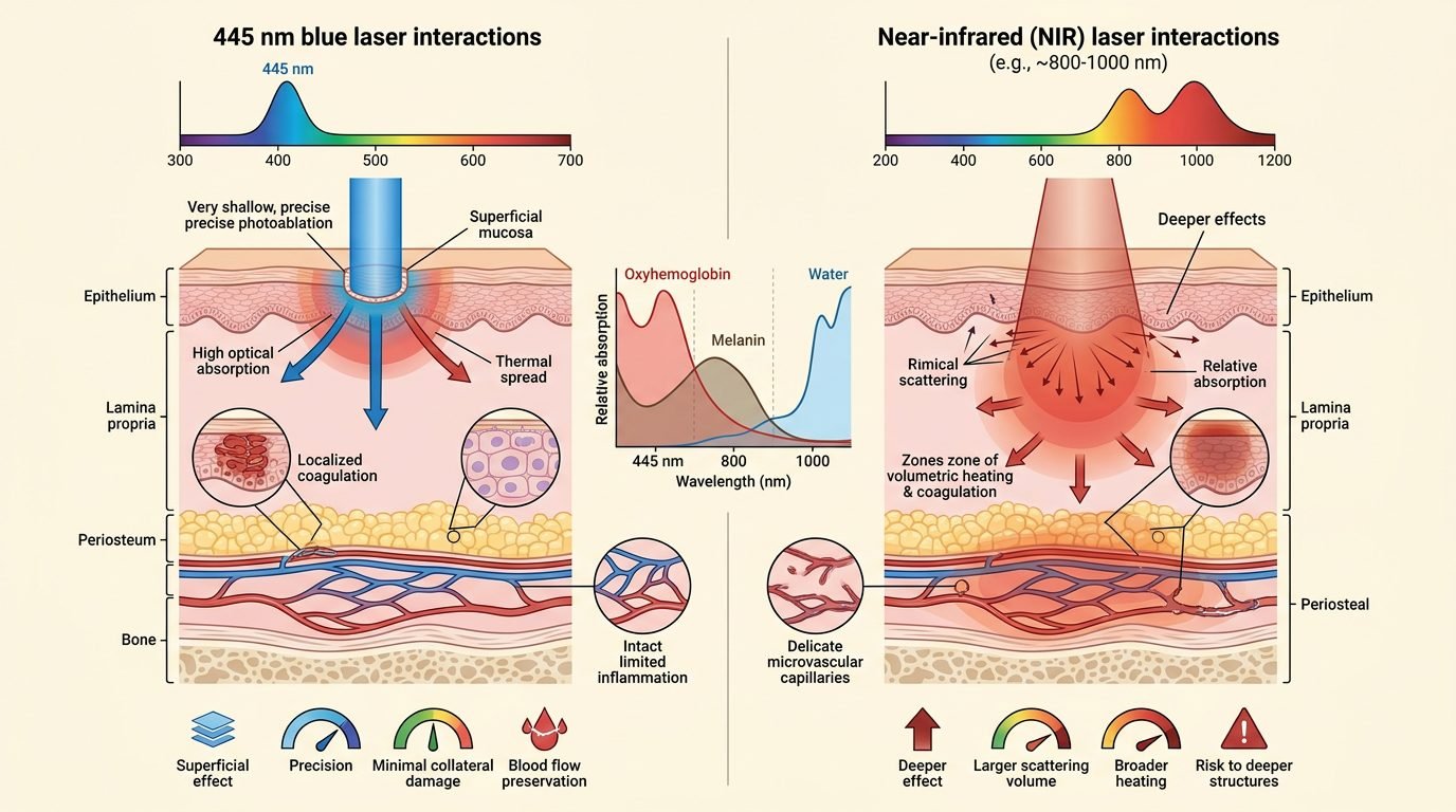
Parameter calibration defines the line between precise epithelial ablation and deep connective tissue burns. Clinicians must utilize non-contact handpieces or cleaved optical fibers (three hundred to four hundred micrometers in diameter) held perpendicular to the tissue surface at a fixed distance of one to two millimeters. Continuous wave (CW) mode should be avoided in favor of pulsed or chopped emission modes to allow adequate thermal relaxation between energy pulses. Clinical studies show that an average power setting between 0.6 and 1.2 watts delivered with pulse durations of ten milliseconds and duty cycles of thirty to fifty percent achieves rapid vaporization of the epithelial stratum spinosum and stratum basale. High-volume intraoral evacuation and a gentle stream of filtered ambient air dissipate surface heat and remove the microscopic ablation plume. Water spray should not be utilized during blue laser de-epithelialization because pooled surface water scatters the collimated blue optical beam and impedes direct visual assessment of mucosal layer clearance.
Clinical sequencing requires methodical visual monitoring of tissue color transitions during de-epithelialization. Following local infiltration anesthesia without excessive vasoconstrictor distension, the clinician moves the blue laser fiber in light, overlapping circular strokes across the designated peri-implant recipient area. The non-keratinized movable mucosa initially turns chalky white as cellular proteins coagulate, followed by gentle layer-by-layer sublimation. De-epithelialization is clinically complete when the superficial epithelial layer is removed, revealing the pale, glistening pink surface of the dense underlying lamina propria and periosteum without exposing yellow submucosal fat globules or white cortical bone plates. Once the bed is prepared, the freshly harvested autogenous graft or acellular dermal matrix (ADM) is positioned immediately, stabilized with resorbable periosteal sutures, and secured with compressive cross-mattress sling sutures to maintain uninterrupted tissue contact.
Longitudinal prospective clinical trials evaluating peri-implant soft tissue augmentation confirm that recipient beds prepared with 445 nm blue lasers achieve postoperative keratinized tissue widths and mucosal thickness values entirely comparable to classic cold-blade surgical scalpel techniques. Furthermore, patients report significantly lower post-operative pain scores, reduced analgesic consumption, and minimal swelling during the initial seven days. The bloodless surgical field dramatically reduces total operative chair time, eliminates suturing interference from active capillary bleeding, and ensures predictable immobilization of delicate connective tissue grafts.
Biochemical and immunohistochemical analyses provide deeper insight into post-ablation tissue healing dynamics. Studies tracking vascular endothelial growth factor (VEGF) secretion and CD31-positive endothelial cell density demonstrate that tissues conditioned with blue semiconductor wavelengths initiate early capillary budding forty-eight hours faster than beds prepared with rotary instrumentation. Mechanical rotary burs produce frictional shear stress that micro-fractures the superficial periosteal collagen framework, generating a thick necrotic smear layer that slows fibroblastic attachment. In contrast, 445 nm photo-ablation creates an ultra-smooth, sterile proteinaceous interface that enhances initial blood clot adhesion and promotes rapid migration of host multipotent mesenchymal stem cells into the graft matrix. This enhanced biological integration correlates directly with long-term peri-implant marginal bone stability: maintaining thick, immobile keratinized mucosa prevents apically directed bacterial penetration, stabilizing crestal bone levels over multi-year follow-up intervals.
Optical biosafety demands non-negotiable operatory protocols. The blue laser beam presents severe ocular hazards due to high transmission through the clear ocular media and focused absorption on the human retina, capable of inducing permanent macular photocoagulation scotomas. Every individual in the operatory, including the patient, clinician, and surgical assistants, must wear certified wavelength-specific protective eyewear possessing an optical density rating of OD 5+ at 445 nm. Reflective metal surgical instruments such as retractors and mouth mirrors must feature non-reflective matte or ceramic coatings to prevent accidental specular reflection of the high-intensity blue beam onto neighboring non-target tissues.
Regarding peri-implant plastic surgery, the 445 nm blue diode laser represents an evidence-supported, micro-precise surgical instrument for soft tissue recipient bed preparation. Its high absorption in primary biological chromophores allows targeted epithelial removal while preserving the vital periosteal vascular network necessary for autograft survival. When operated within conservative wattage limits, pulsed emission cycles, and strict ocular safety standards, blue laser de-epithelialization minimizes intraoperative trauma, accelerates plasmatic graft integration, and ensures stable, long-term keratinized mucosal architecture around dental implants.
Leave a Reply