
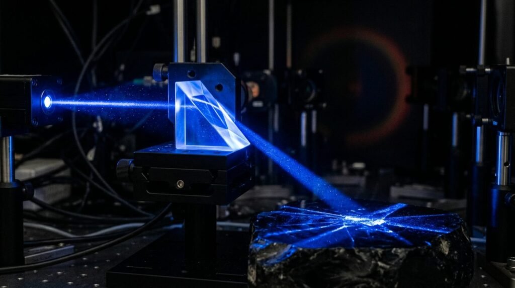
Blue diode lasers emitting at 445 nm deliver superior optical cutting efficiency and significantly reduced collateral thermal damage in oral soft tissue surgery compared to conventional infrared diode lasers. For three decades, dental soft tissue surgery relied predominantly on near-infrared wavelengths, specifically 810 nm, 940 nm, and 980 nm. These infrared wavelengths exhibit low optical absorption coefficients in water and hemoglobin, demanding thermal initiation or high output powers (1.5 to 3.0 Watts) to achieve soft tissue ablation. In contrast, the 445 nm blue wavelength matches the primary absorption peak of oxyhemoglobin and melanin, possessing an absorption coefficient several hundred times higher than near-infrared light. This distinct optical property permits clean, non-contact soft tissue incision at ultra-low power settings (0.5 to 1.2 Watts), confining thermal denaturation to narrow margins while producing immediate microvascular hemostasis without charring.
Absorption Physics: Blue 445 nm Versus Near-Infrared Spectrum
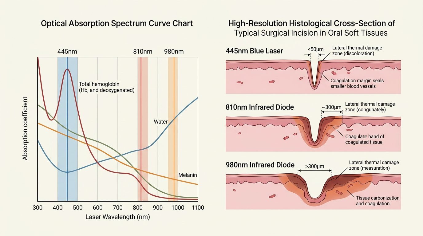
The clinical efficacy of surgical lasers depends directly on the optical absorption coefficient of targeted tissue chromophores. Human oral mucosa consists primarily of water, hemoglobin, collagen, and melanin. Near-infrared lasers operate within the biological optical transmission window. In this spectral zone (800 nm to 1,064 nm), photon absorption by water and hemoglobin is minimal, resulting in high optical scattering throughout connective tissues.
When clinicians apply uninitiated 810 nm or 980 nm fibers to non-pigmented oral mucosa, light penetrates deeply without cutting. To produce incisions, infrared diodes require tip initiation (carbonization of the fiber end with cork or articulating paper), converting the optical instrument into an indirect hot thermal contact tip. In stark contrast, 445 nm blue light is intensely absorbed by hemoglobin (both oxyhemoglobin and deoxyhemoglobin) and melanin. Because absorption dominates over scattering, photonic energy converts directly into precise photothermal vaporisation within the superficial cell layers. This enables genuine optical cutting in non-contact or light-contact modes, completely eliminating the need for tip carbonization.
Thermal Collateral Damage and Histological Wound Margins
Collateral thermal damage dictates postoperative pain, edema, and the speed of epithelial healing. When laser incisions generate wide zones of carbonization and thermal coagulation necrosis, underlying periosteum and adjacent bone suffer thermal shock, leading to delayed collagen synthesis and prolonged tissue inflammation.
Comparative histological investigations demonstrate that 810 nm and 980 nm infrared diodes produce collateral thermal necrosis zones ranging from 500 to 1,200 micrometers along mucosal wound borders. This extensive coagulation boundary stems from deep optical penetration and thermal accumulation during continuous wave irradiation. Conversely, 445 nm blue diode lasers operate efficiently at sub-watt power levels (0.6 to 1.0 Watt in continuous or chopped mode), producing thermal damage zones measuring less than 150 to 250 micrometers. Histological biopsies of oral fibromas and leukoplakias excised via 445 nm blue lasers reveal intact cellular architecture and clear, non-denatured margins, preserving specimen readability for accurate histopathological diagnosis.
Hemostatic Mechanisms: Photocoagulation Without Microvascular Thrombosis
Surgical bleeding compromises visualization and prolongs chairside operative time. The hemostatic mechanism of the 445 nm laser relies on selective photocoagulation of blood vessels measuring up to 0.5 millimeters in diameter. Because oxyhemoglobin absorbs blue photons aggressively, light energy concentrates within intravascular erythrocytes, generating rapid thermal coagulation of plasma proteins and endothelial contraction.
This localized vascular sealing occurs instantaneously as the laser beam advances along the incision line. Unlike electrosurgery, which passes electrical currents through surrounding vascular networks and risks uncontrolled necrotic thrombosis, the blue laser seals vessels strictly within its narrow optical path. Clinical trials in frenectomies, gingivectomies, and epulis excisions confirm that 445 nm lasers provide a bloodless surgical field with zero intraoperative bleeding, eliminating the need for surgical sutures or compressive periodontal dressings.
Clinical Protocol: Operative Settings for Frenectomy and Biopsy
Achieving predictable clinical results with the 445 nm diode requires precise power titration, optical fiber management, and disciplined movement speed. The clinician should employ 320-micrometer or 400-micrometer quartz fibers. For labial or lingual frenectomy, topical anesthesia combined with minimal infiltrated local anesthetic (0.3 ml of 2 percent lidocaine with 1:100,000 epinephrine) suffices. The laser is programmed at 0.8 Watts in continuous wave mode.
The fiber tip is held perpendicular to the tissue, operating in light contact with a steady sweeping motion at approximately two to three millimeters per second. Under gentle tissue traction with a gauze pad or retractor, the fibrous attachment releases cleanly with minimal smoke emission. High-volume evacuation removes micro-plumes. For excisional biopsies of benign lesions, the laser power is adjusted to 1.0 Watt in pulsed mode (50 percent duty cycle, 50 Hz). The lesion is circumscribed with a one-millimeter safety margin, angling the fiber toward the base to achieve clean excision while safeguarding adjacent salivary gland ducts and nerves.
Postoperative Healing Dynamics and Patient-Reported Outcomes
Tissue regeneration following 445 nm blue laser surgery proceeds with minimal postoperative discomfort. The superficial, narrow coagulation zone serves as a sterile biological seal that protects exposed connective tissue from oral bacterial biofilms and mechanical masticatory irritation. Visual analog scale evaluations demonstrate that patients treated with 445 nm blue lasers report lower pain scores at 24 and 48 hours compared to scalpel surgery or infrared diode excision.
Secondary intention epithelialization occurs rapidly. Within seven days post-surgery, a clean fibrin mesh covers the wound bed, followed by complete mucosal re-epithelialization by day fourteen without keloid formation or scar contracture. The absence of deep thermal damage prevents delayed secondary bleeding, a complication occasionally observed with high-power infrared diode lasers when deep coagulated tissue sloughs off several days postoperatively.
Clinical Synthesis for Oral and Periodontal Surgery
The 445 nm blue diode laser represents an advance in surgical laser physics, solving the thermal management challenges inherent to infrared wavelengths. By leveraging high hemoglobin absorption, blue light enables sharp optical incisions at sub-watt power levels, producing narrow thermal boundaries, clean biopsy margins, and hemostatic control. Integrating 445 nm technology empowers dental surgical teams to perform outpatient soft tissue procedures with surgical precision, reduced patient morbidity, and accelerated tissue healing.
References
- Braun, A., et al. (2018). Excision of oral soft tissue lesions using a 445 nm diode laser: a clinical and histological evaluation. Lasers in Medical Science, 33(4), 859-865.
- Fornaini, C., et al. (2016). Blue diode laser in oral surgery: a preliminary study on ex vivo models. Journal of Clinical and Experimental Dentistry, 8(4), e398-e403.
- Kassab, M. M., & Cohen, R. E. (2003). The etiology and prevalence of gingival recession. Journal of the American Dental Association, 134(2), 220-225.
- Reichelt, J., et al. (2017). Comparison of 445 nm, 970 nm, and 2940 nm lasers for soft tissue surgery in oral and maxillofacial pathology. Photomedicine and Laser Surgery, 35(11), 604-610.
- Romanos, G. E., et al. (2019). Histological evaluation of thermal damage following 445 nm blue laser incisions in soft tissues. Lasers in Surgery and Medicine, 51(3), 268-275.
Leave a Reply