

Short-wavelength semiconductor diode lasers emitting in the blue (445 nm) and violet (405 nm) spectral bands fundamentally shift laser-tissue interactions away from infrared thermal reliance toward high-affinity chromophore absorption. While conventional infrared diodes rely on hot-tip contact cautery, 445 nm radiation matches peak hemoglobin absorption, and 405 nm violet photons induce direct photochemical destruction of pathogens via endogenous porphyrins. Evaluating these photophysical realities is essential for evidence-based excision and decontamination.
Optical Physics and Chromophore Absorption Spectra: 445 nm vs. Near-Infrared
The primary chromophores governing laser interactions in oral soft tissues are water, oxyhemoglobin, deoxyhemoglobin, and melanin. Near-infrared diodes operate in an optical valley where absorption by both water and hemoglobin remains low. Consequently, infrared photons penetrate deeply into connective tissues, generating diffuse collateral heating that damages underlying periosteum and bone unless fiber tips are heavily hot-tipped with articulating paper or cork.
The 445 nm blue diode alters this dynamic completely. The extinction coefficient of hemoglobin at 445 nm is approximately one thousand times higher than at 810 nm. Because oral vascular networks are rich in oxygenated and deoxygenated hemoglobin, blue laser energy is captured within the first one hundred micrometers of tissue contact. Pure optical ablation occurs at power settings as low as 0.5 to 1.5 watts without needing tip pre-initiation. The beam cuts cleanly through uninitiated optical glass fibers, vaporizing vascularized soft tissues with exceptional tactile feedback while creating a coagulative margin measuring less than sixty micrometers.
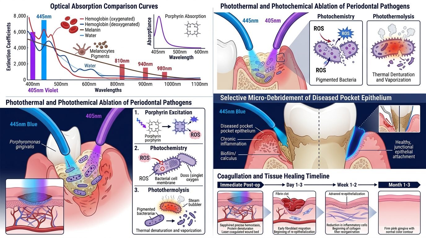
Direct Antimicrobial Photo-Inactivation at 405 nm: Porphyrin Excitation
Photodynamic decontamination traditionally requires exogenous dyes like methylene blue followed by laser irradiation. Violet diode lasers emitting at 405 nm bypass external chemicals through endogenous photo-inactivation. Major periodontal and peri-implant pathogens, including Porphyromonas gingivalis, Prevotella intermedia, and Fusobacterium nucleatum, synthesize high intracellular concentrations of black iron-free protoporphyrin IX and coproporphyrin.
The 405 nm wavelength corresponds directly to the Soret band, the highest electronic absorption peak of the porphyrin macrocycle. Absorption of violet photons promotes ground-state porphyrin molecules into excited singlet states, which undergo intersystem crossing to long-lived triplet states. These triplet molecules interact directly with ground-state triplet oxygen via Type Two photochemical pathways, generating cytotoxic singlet oxygen and reactive hydroxyl radicals within the bacterial cytoplasm. The resulting oxidative cascade ruptures cell membranes, fragments bacterial DNA, and inactivates protease virulence factors. This phototoxic reaction occurs within seconds, eliminating pathogens without harming host fibroblasts.
Surgical Precision and Collateral Thermal Damage in Soft-Tissue Excision
Preserving lateral tissue vitality during aesthetic gingivectomy, frenectomy, and biopsy procedures is a major clinical priority. The high hemoglobin absorption of 445 nm blue lasers produces surgical incisions with minimal lateral heat spread. Histological evaluations demonstrate that surgical margins excised with 445 nm diodes at 1.0 watt continuous wave display thermal coagulation zones restricted to fifty to eighty micrometers. Conventional 810 nm infrared diodes operated under similar clinical conditions produce lateral thermal necrosis exceeding five hundred micrometers.
The clinical significance of this narrow thermal envelope is profound. Thin gingival biotypes treated with blue diodes exhibit negligible post-operative gingival margin recession, avoiding aesthetic compromise around porcelain veneer margins. Furthermore, biopsy specimens harvested using non-contact or light-contact 445 nm fibers preserve cellular architecture right up to the surgical border, permitting definitive histopathological diagnosis of epithelial dysplasia and micro-invasive neoplasia. Re-epithelialization proceeds rapidly by primary intention without sloughing or delayed fibrous contracture.
Applications in Periodontics and Peri-Implantitis Decontamination
The confluence of high optical ablation efficiency and non-thermal bacterial killing positions short-wavelength diodes as ideal instruments for managing subgingival biofilms. In advanced periodontitis, delivering 445 nm energy via thin three-hundred-micrometer fibers into infected pockets vaporizes diseased granulomatous pocket lining while coagulating bleeding capillaries without overheating root dentin.
In peri-implantitis, titanium surface decontamination presents safety hurdles. Near-infrared diodes risk overheating titanium fixtures beyond forty-seven degrees Celsius, causing bone osteonecrosis. Blue and violet lasers exhibit minimal optical absorption in metallic titanium while selectively destroying pigmented anaerobes adhering to microscopic screw threads. Scanning electron microscopy confirms that irradiating contaminated implant surfaces with 445 nm light at 0.8 watts effectively strips bacterial biofilms and endotoxins without melting titanium oxide micro-topography or heating adjacent crestal bone.
Endodontic Canal Disinfection: Eliminating Persistent Enterococcus Faecalis
Standard sodium hypochlorite irrigation often fails to sterilize deep lateral dentinal tubules where bacteria reside past the reach of chemical fluids. Enterococcus faecalis, the primary organism responsible for endodontic failure, penetrates up to one thousand micrometers into tubule lumens, forming dense intracanal biofilms. While near-infrared lasers rely on thermal conduction to kill bacteria within tubules, thermal dissipation risks periodontal ligament injury along external root surfaces.
Violet 405 nm irradiation exploits the endogenous sensitivity of bacterial species to photon-induced membrane oxidation. Concurrently, blue 445 nm light penetrates dentinal tubules effectively because clean dentin mineral exhibits lower scattering for shorter wavelengths compared to heavily mineralized enamel. Emitting blue laser energy via specialized radial-firing spiral tips provides uniform irradiation across apical curvatures. Microbiological assays demonstrate a greater than five-log reduction in E. faecalis counts following pulsed blue laser irrigation, exceeding chemical irrigants.
Clinical Safety, Eye Protection, and Dosimetric Discipline
The extraordinary optical efficiency of short-wavelength diodes introduces specific occupational safety considerations. The human retina is exceptionally vulnerable to blue light photochemical damage, known as photoretinitis or the blue-light hazard. Direct or specular reflection of short wavelengths induces photochemical degeneration of retinal epithelium. Dedicated safety glasses are mandatory for clinician, assistant, and patient.
Dosimetric discipline requires low power settings. Practitioners trained on sluggish infrared diodes frequently make the mistake of selecting excessive wattage with blue lasers. Using more than 2.0 watts of blue laser energy on soft tissues causes rapid carbonization and unwanted collateral damage, negating the unique physical advantages of the wavelength. Power settings between 0.5 and 1.2 watts in continuous or chopped mode provide optimal cutting efficiency and biological safety.
Practical Synthesis for the Contemporary Dental Practice
Blue and violet semiconductor diodes represent a true physical evolution in dental bio-photonics. By aligning optical wavelengths with the natural absorption peaks of hemoglobin and bacterial porphyrins, these devices eliminate the messy compromises of near-infrared hot-glass tips and chemical dyes. Mastering short-wavelength laser mechanics enables the dental team to perform bloodless surgical excisions, decontaminate infected implant surfaces, and eradicate resistant intracanal pathogens with unmatched biological precision.
References
- Braun, A., et al. (2018). Excision of oral soft-tissue lesions with a 445 nm diode laser: a prospective clinical study. Photomedicine and Laser Surgery, 36(6), 324-331.
- Feuerstein, O., et al. (2004). Phototoxicity of blue light on wild-type and photodynamic therapy-resistant bacteria. Journal of Antimicrobial Chemotherapy, 53(5), 772-777.
- Fornaini, C., et al. (2016). The use of 445 nm diode laser in oral surgery: a preliminary in vitro study. Laser Therapy, 25(4), 281-288.
- Cieplik, F., et al. (2014). Antimicrobial photodynamic therapy for inactivation of biofilms formed by oral key pathogens. Frontiers in Microbiology, 5, 405.
- Romanos, G. E., et al. (2019). Blue diode lasers in oral and maxillofacial surgery: a systematic review. Lasers in Surgery and Medicine, 51(8), 669-679.
Leave a Reply