
The carbon dioxide laser operating at 10,600 nanometers provides unsurpassed precision in oral soft tissue surgery through extraordinary absorption in tissue water, creating rapid vaporization with minimal collateral thermal necrosis. Oral soft tissue interventions frequently challenge clinicians due to high vascularity, salivary contamination, and complex muscular dynamics in anatomical zones such as the tongue, floor of the mouth, and buccal mucosa. Conventional scalpel excision induces intraoperative hemorrhage that impairs visibility, requires sutures, and produces significant postoperative myofascial contraction. Conversely, electrosurgery and non-contact diode lasers often generate wide thermal artifacts that compromise biopsy margins and delay re-epithelialization. The 10,600 nm carbon dioxide (CO2) laser resolves these clinical dilemmas by combining immediate microvascular coagulation with a tightly controlled thermal damage zone under 200 micrometers.
Optical Physics: Water Absorption Peak and Thermal Confinement
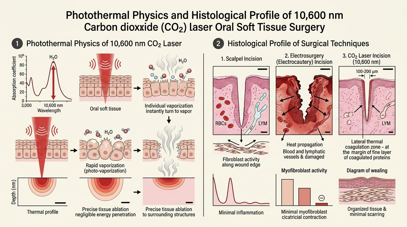
The operative efficacy of the CO2 laser derives from its interaction with intracellular and extracellular water molecules. With an emission wavelength in the mid-infrared region (10,600 nm), its absorption coefficient in liquid water reaches approximately 800 to 1000 cm-1. Because oral epithelium and stroma contain 75% to 85% water, the incident radiant energy is absorbed within the superficial 10 to 30 micrometers of tissue.
This localized energy deposition causes rapid boiling of intracellular water, leading to explosive cellular rupture via photothermal vaporization. Operating in superpulsed or ultrapulsed modes prevents thermal diffusion into adjacent structures. When pulse duration remains shorter than the thermal relaxation time of oral soft tissue (600 to 800 microseconds), thermal energy cannot conduct into deep layers. The resulting lateral zone of thermal coagulation measures between 50 and 150 micrometers. This narrow margin seals microcapillaries, terminal lymphatic vessels, and sensory nerve endings while preserving neighboring cellular architecture.
Hemostatic Dynamics and Blood Vessel Coagulation Limits
Intraoperative hemostasis during CO2 laser excision relies on thermal denaturing of collagen and plasma proteins within vessel walls. The lateral heat zone generates temperatures between 60 and 100 degrees Celsius, inducing immediate microvascular occlusion. Capillaries, venules, and arterioles up to 0.5 millimeters in diameter are coagulated instantaneously during incision.
This dry surgical field provides clear visualization of anatomical margins, eliminating suctioning required during cold blade surgery. However, clinicians must recognize anatomical limitations: vessels larger than 0.6 millimeters, such as branches of the lingual or facial artery, cannot be reliably sealed by radiant energy alone. In deep lingual dissections, surgical clips or sutures remain necessary to secure large pulsatile vessels. The coagulation of unmyelinated sensory C-fibers and delta-A nerve endings blocks noxious depolarization, accounting for the marked reduction in acute postoperative discomfort reported by patients.
Surgical Biopsy: Preserving Histological Margins
Performing diagnostic excisional biopsies with laser energy requires rigorous technique to avoid thermal artifacts that complicate histopathological interpretation. When an uncalibrated laser beam or excessive energy density is directed across a biopsy margin, thermal coagulation produces cell elongation, nuclear pyknosis, and cytoplasmic vacuolization. Pathologists describe this artifact as “streaming,” which can mask dysplasia or simulate invasive squamous cell carcinoma.
To preserve diagnostic integrity, clinicians must adopt specific surgical guidelines when excising suspicious leukoplakias, erythroplakias, or mucosal nodules. A superpulsed mode should be selected with a narrow focal spot diameter (0.2 to 0.4 mm) and power restricted to 4 to 6 Watts. The incision must maintain a safety margin of at least two millimeters beyond the clinical lesion boundary. The beam must be kept in rapid, continuous motion, perpendicular to the mucosal surface, avoiding hovering over a single point. Histopathological studies confirm that adherence to these parameters restricts marginal thermal artifacts to under 100 micrometers, allowing unequivocal evaluation of surgical margins.
Ablation and Vaporization of Extensive Premalignant Lesions
For extensive, multifocal leukoplakias or lichen planus with verrucous proliferation covering wide areas of the buccal mucosa or hard palate, surgical resection with primary closure causes severe scar contracture and limited mouth opening. In these scenarios, CO2 laser vaporization provides tissue preservation by stripping the pathological epithelial layer without removing underlying submucosal muscle beds.
Clinicians defocus the handpiece by increasing working distance, enlarging the focal spot to 1.0 to 1.5 millimeters. At a power setting of 4 to 8 Watts, the beam sweeps across the lesion like a brush. The atypical epithelial layer is converted to vapor layer-by-layer until healthy, non-keratinized basement membrane and fibrous stroma are revealed. The surface is wiped with sterile moist gauze to inspect residual lesion islands. Because the underlying elastic lamina and deep collagen beds remain intact, the surgical site heals without cicatricial tethering or microstomia.
Wound Healing Dynamics and Secondary Epithelialization
The biological healing cascade following CO2 laser surgery differs fundamentally from scalpel wounds. Scalpel incisions closed with sutures heal by primary intention, whereas CO2 laser wounds are typically left unsutured to heal by secondary intention under a biological coagulum. The thermal denaturation layer forms an insoluble surface protein seal that protects the surgical bed from oral bacterial colonization, mechanical salivary shearing, and food debris irritation.
Histological studies demonstrate an inflammatory delay of 48 to 72 hours compared to scalpel cuts. Polymorphonuclear leukocyte infiltration is minimized because open blood vessels are absent. By day five, active granulation tissue emerges from viable lateral margins. Epithelial migration proceeds smoothly across the denatured collagen matrix. Myofibroblastic differentiation is significantly suppressed compared to scalpel wounds; this attenuation of myofibroblast activity accounts for minimal wound contraction and absent hypertrophic scar formation, preserving tongue mobility and mucosal flexibility.
Clinical Protocols and Safety Requirements
Achieving predictable clinical results demands strict environmental and patient safety protocols. The CO2 wavelength presents significant ocular hazards: the cornea absorbs 10,600 nm photons instantly, predisposing to corneal burns and permanent opacification. Every person in the surgical operatory must wear certified protective eyewear with optical density (OD) > 5 at 10,600 nm. Clear plastic or glass side-shielded glasses provide effective barrier protection.
Furthermore, because the laser operates via photothermal vaporization, it generates an aerosolized laser plume containing cellular debris, carbon particles, viral particles, and volatile organic compounds. Clinicians must utilize dedicated high-volume evacuation systems featuring ultra-low particulate air (ULPA) filtration situated within 15 millimeters of the ablation site. Anesthetic endotracheal tubes must be laser-resistant or protected.
References
- Bornstein, M. M., et al. (2005). Comparison of mucosal wound healing after carbon dioxide laser surgery and conventional scalpel surgery: a histological study. Journal of Oral and Maxillofacial Surgery, 63(6), 784-791.
- Fisher, S. E., et al. (1983). The effect of the carbon dioxide surgical laser on oral tissues. The British Journal of Oral & Maxillofacial Surgery, 21(3), 174-185.
- Ishii, J., et al. (2012). Carbon dioxide laser surgery for premalignant and malignant lesions of the oral mucosa: therapeutic outcomes and recurrence factors. Journal of Dental Research, 91(7_suppl), 53S-58S.
- Suter, V. G., et al. (2010). Efficacy and safety of the carbon dioxide laser for oral soft tissue surgeries: a prospective clinical study of 120 patients. Lasers in Medical Science, 25(3), 361-367.
- van der Hem, P. S., et al. (2005). The results of CO2 laser vaporization of oral leukoplakia with a minimum follow-up of 5 years. Oral Oncology, 41(1), 31-37.
Leave a Reply