
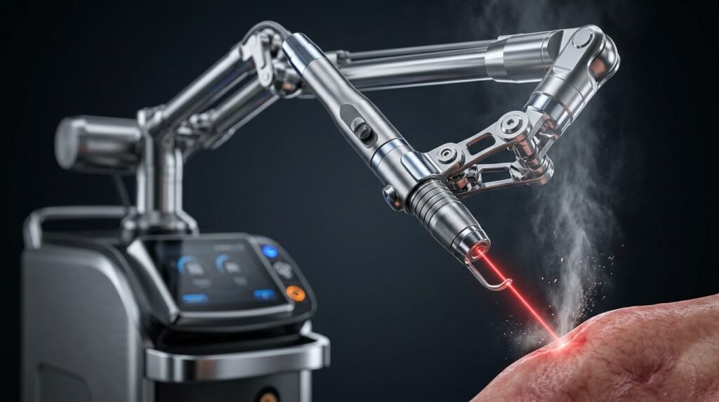
Oral leukoplakia represents the most prevalent potentially malignant disorder affecting the oral cavity, carrying documented transformation rates into invasive squamous cell carcinoma ranging between one and twenty percent. Management requires eradicating dysplastic mucosal epithelium while minimizing postoperative morbidity, scarring, and functional tongue impairment. Conventional scalpel excision produces tissue loss, dense scar contracture, and high recurrence rates exceeding thirty-five percent due to field cancerization. Carbon dioxide laser systems, operating at 10,600 nanometers and 9,300 nanometers, provide an ideal biophysical balance of high water absorption, shallow optical penetration, and continuous microvascular coagulation. Photothermal vaporization or excisional biopsy using focused carbon dioxide beams achieves precise cellular ablation of epithelial dysplasia, preserves deep muscular architectures, seals mucosal lymphatic vessels, and reduces recurrence and malignant transformation rates compared to conventional surgical scalpels.
Photobiophysics of Carbon Dioxide Radiation and Water Absorption
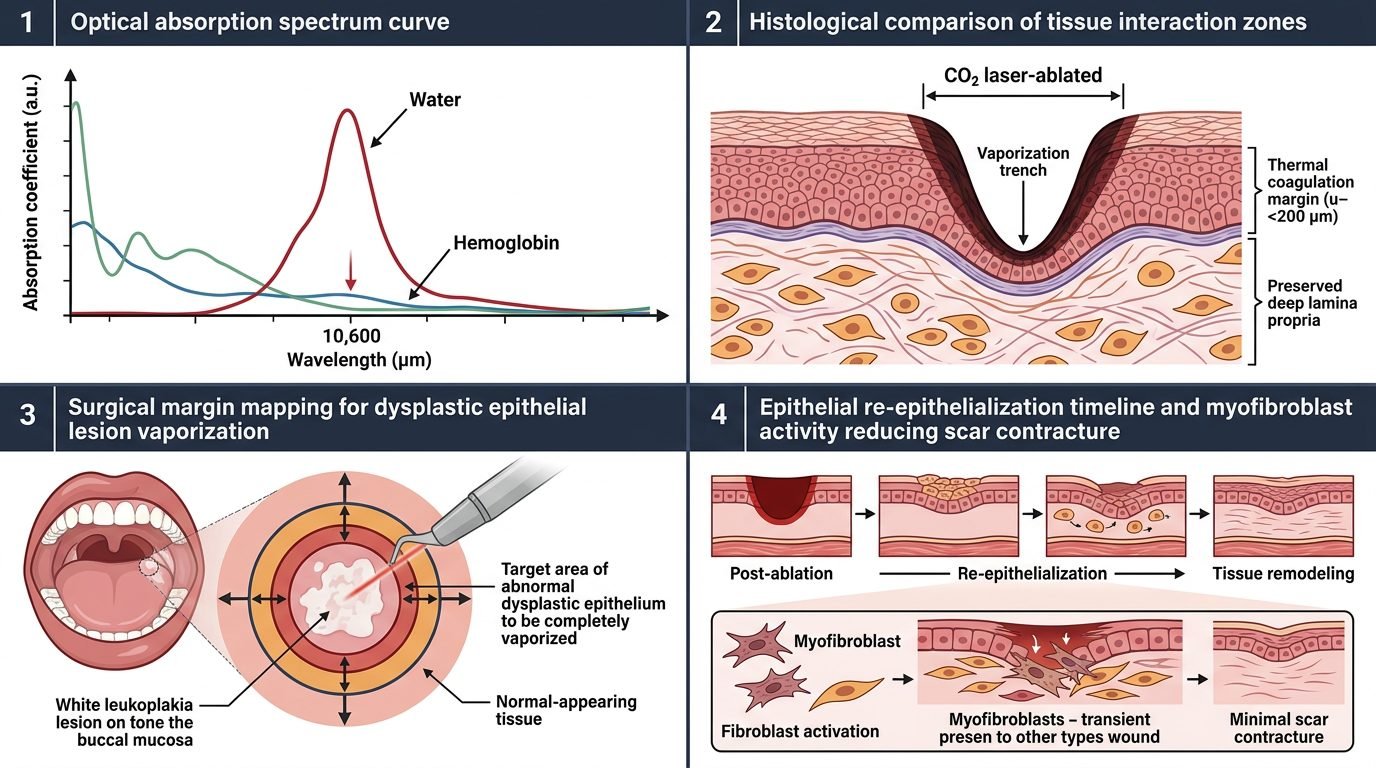
The carbon dioxide laser delivers infrared electromagnetic energy at 10,600 nm, coinciding directly with the fundamental vibrational resonance mode of biological water molecules. Biological oral mucosa consists of over seventy-five percent intracellular and extracellular water. Consequently, the absorption coefficient of oral soft tissues at 10,600 nm is exceptionally high, resulting in an optical penetration depth restricted to approximately twenty to thirty micrometers.
When the focused carbon dioxide laser beam interacts with dysplastic epithelial tissue, photonic energy is absorbed almost instantaneously by intracellular water. Instantaneous boiling occurs within microseconds, converting liquid water into expanding micro-steam bubbles that burst cellular membranes through explosive volumetric expansion. This localized micro-explosion produces immediate cellular vaporization. Because energy conversion is rapid and shallow, collateral thermal damage extends laterally by only one hundred to two hundred micrometers, leaving underlying deep muscular stroma and lamina propria structurally undamaged.
Field Cancerization and the Limitations of Cold-Blade Excision
The primary clinical dilemma in leukoplakia eradication is the phenomenon of mucosal field cancerization. Chronic mucosal exposure to carcinogens, including tobacco nitrosamines, alcohol metabolites, and betel quid, induces widespread genetic alterations throughout clinically normal mucosal sectors surrounding the visual leukoplakic plaque. Keratinocytes in these adjacent zones frequently exhibit loss of heterozygosity, p53 tumor suppressor gene mutations, and chromosomal aneuploidy despite displaying completely benign histological architecture on biopsy.
When a surgeon attempts scalpel excision, achieving clear margins requires wide peripheral tissue sacrifice that compromises speech, swallowing, and salivation. If margins are kept narrow to preserve function, genetically transformed clone cells remain behind in the surgical bed, inevitably driving lesion recurrence or de novo malignant transformation. Conversely, carbon dioxide laser vaporization permits generous peripheral sweeping across a three-to-five-millimeter safety margin of surrounding mucosa. The thermal plume ablates superficial micro-foci of clonally expanded dysplastic cells with zero blood loss, neutralizing subclinical field cancerization without inducing functional mutilation.
Microvascular Coagulation and Lymphatic Sealing Mechanisms
A distinctive surgical attribute of carbon dioxide laser vaporization is instantaneous coagulation of microvascular and lymphatic networks. The zone of collateral thermal denaturation (one hundred to two hundred micrometers) heats local capillary walls to approximately one hundred degrees Celsius. This controlled temperature rise denatures vascular endothelial proteins and activates plasma fibrin cross-linking, sealing capillaries and venules up to 0.5 millimeters in diameter.
Simultaneously, carbon dioxide laser irradiation seals severed lymphatic channels. In the event that an undiagnosed micro-invasive squamous cell focus resides within a dense leukoplakic plaque, sealing lymphatic vessels during excision prevents iatrogenic intravascular dissemination of malignant tumor cells into cervical lymph nodes. The resulting surgical field remains completely dry and pristine, providing the clinician with unobstructed visual control over dysplastic tissue margins throughout the operative procedure.
Epithelial Regeneration, Myofibroblasts, and Scarless Healing
Mucosal wound healing following carbon dioxide laser vaporization proceeds through distinct biological pathways compared to mechanical scalpel incisions. Secondary-intention healing of laser-ablated mucosal defects produces exceptionally little wound contracture. Histological and immunohistochemical evaluations reveal that carbon dioxide laser wounds exhibit delayed and diminished infiltration of alpha-smooth muscle actin-positive myofibroblasts.
Because myofibroblastic differentiation is subdued, the physical contraction forces that typically pull wound margins together are absent. Instead, the surgical site is covered by a natural biological dressing of coagulated denatured plasma protein that shields exposed sensory nerve endings from mechanical friction and oral fluids. Within seven days, epithelial keratinocytes migrate smoothly across the wound bed beneath the coagulum. By four weeks, complete re-epithelialization is achieved with pliable, soft tissue that preserves full tongue flexibility and avoids painful tethering or restrictive scarring.
Clinical Dosimetry and Operatory Ablation Protocols
Executing predictable carbon dioxide laser ablation of oral leukoplakia demands rigorous dosimetric control. For diagnostic safety, any heterogeneous leukoplakia displaying verrucous, nodular, or erythematous components (erythroleukoplakia) must undergo an initial laser excisional biopsy rather than pure vaporization to allow comprehensive histological grading of epithelial dysplasia.
For homogeneous lesions undergoing definitive vaporization, the carbon dioxide laser is configured in continuous-wave or super-pulsed mode with a spot diameter of 0.8 to 1.0 millimeters. Power output is maintained between 4 and 8 watts, delivering an energy density of approximately 10 to 15 Joules per square centimeter. The handpiece sweeps in systematic overlapping cross-hatch passes, vaporizing dysplastic epithelium layer by layer until the healthy, glistening fibrous architecture of the lamina propria is reached. A moist sterile gauze clears carbonized coagulum between passes, preventing heat accumulation and ensuring precise visual depth verification.
Long-Term Recurrence Profiles and Malignant Transformation
Longitudinal clinical trials monitoring hundreds of patients over five-to-ten-year follow-up intervals demonstrate that carbon dioxide laser ablation achieves recurrence rates between ten and eighteen percent, representing a dramatic improvement over the thirty-five to forty percent recurrence observed after conventional scalpel excision. More importantly, the rate of malignant transformation into oral squamous cell carcinoma drops from approximately twelve percent in scalpel-treated cohorts to under five percent following comprehensive laser vaporization.
Because laser vaporization induces negligible scarring, repeat interventions for recurrent dysplastic plaques can be executed multiple times without compounding structural deformities or functional limitations. Regular three-month clinical surveillance coupled with photographic documentation and vital staining ensures lifelong oral mucosal health.
Significance in Modern Oral Oncology and Preventative Care
Carbon dioxide laser vaporization represents a cornerstone modality in preventative oral oncology. By exploiting precise infrared water absorption to eradicate dysplastic epithelium while neutralizing microscopic field cancerization, clinicians safeguard mucosal architecture and prevent malignant progression. Laser precision elevates patient quality of life, replacing invasive mutilating resections with conservative, highly effective biophysical preservation.
References
- van der Hem, P. S., et al. (2005). CO2 laser evaporation of oral leukoplakia: a retrospective study of 200 cases. Oral Oncology, 41(5), 488-494.
- Ishii, J., et al. (2004). Assessment of CO2 laser surgery for oral leukoplakia: a study of 42 patients. Journal of Oral and Maxillofacial Surgery, 62(10), 1238-1243.
- Brouns, E. R., et al. (2014). Treatment of oral leukoplakia: a review of the literature. Medicina Oral, Patología Oral y Cirugía Bucal, 19(5), e440-e449.
- Chiesa, F., et al. (1993). Follow-up of oral leukoplakia after carbon dioxide laser surgery. Acta Otorhinolaryngologica Italica, 13(5), 421-429.
- Warnakulasuriya, S., et al. (2007). Nomenclature and classification of potentially malignant disorders of the oral mucosa. Journal of Oral Pathology & Medicine, 36(10), 575-580.
Leave a Reply