

Persistent idiopathic orofacial pain exposes the failure of structural reductionism in clinical dentistry. Patients suffering from chronic facial pain, burning sensations, and atypical odontalgia frequently undergo successive invasive interventions without receiving symptom relief. Invasive interventions on sound teeth fail because pathology originates in altered central neuroplasticity and somatosensory amplification rather than peripheral infection. Humanized dental care requires diagnostic restraint and communication strategies that validate suffering without unwarranted surgery.
Neurobiological Mechanisms of Central Sensitization and Somatosensory Amplification
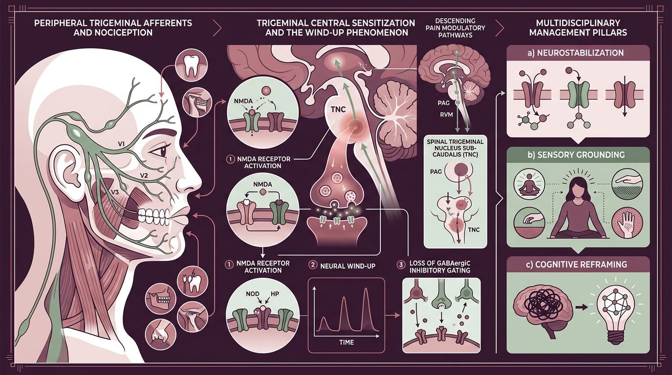
Central sensitization describes an operational state of the nervous system characterized by generalized nociceptive hypersensitivity. Following prolonged peripheral input or surgical trauma, second-order neurons within the spinal trigeminal nucleus caudalis exhibit persistent hyperexcitability. The threshold for action potential generation decreases, receptive fields expand, and low-threshold mechanoreceptors activate nociceptive pathways. Normal stimuli, including mastication, light touch, and air currents, produce severe discomfort.
Somatosensory amplification acts concurrently at higher cortical processing levels. Patients possessing high somatic amplification perceive normal bodily sensations as intense and alarming. Minor occlusal contacts or temperature shifts are registered as structural emergencies. Functional neuroimaging demonstrates aberrant functional connectivity between the anterior insular cortex, anterior cingulate cortex, and prefrontal emotional networks. The trigeminal input is real; it is processed through neurocircuitry that amplifies intensity while stripping inhibitory descending modulation.
The Cycle of Failed Dentoalveolar Interventions and Iatrogenic Harm
The clinical trajectory of patients with somatoform amplification follows a predictable pattern across dental practices. It typically begins with a vague ache in a posterior tooth without objective radiographic pathology. Convinced that pain equates to pulpal necrosis, the practitioner performs an endodontic procedure. The patient experiences brief relief from local anesthesia and placebo effect. Within weeks, however, the discomfort returns with greater intensity or shifts to an adjacent tooth.
Frustrated by failure, the clinician re-treats root canals, undertakes apical surgery, or extracts the tooth. Each intervention introduces fresh tissue trauma and triggers ectopic neuromatous sprouting. This mechanical cycle compounds the central sensitization state. Clinical cohorts demonstrate that more than seventy percent of patients diagnosed with persistent idiopathic dentoalveolar pain underwent multiple irreversible, unnecessary dental interventions prior to neurological consultation. Recognizing when to stop drilling constitutes an essential clinical skill in modern practice.
Alexithymia, Affective Dysregulation, and Somatic Substitution
Alexithymia, defined as the inability to articulate emotional feelings and differentiate them from bodily sensations, is prevalent among chronic pain cohorts. Individuals with high alexithymic traits experience psychological distress primarily through somatic channels. Masseter hypertonicity, autonomic tension, and trigeminal hypersensitivity become the physiological vocabulary through which unexpressed emotional pain is manifested.
When clinicians dismiss these individuals as hysterical or deceptive, the therapeutic relationship ruptures. The patient interprets abrupt psychiatric referrals as an accusation of malingering, prompting defensive entrenchment. The patient doubles down on somatic complaints, searching for another practitioner willing to operate. A humanized clinician recognizes that somatic substitution represents an involuntary protective adaptation. Pain validation must occur at the physiological level before psychological co-factors can be introduced into clinical dialogue.
Clinical Communication Protocols: De-escalation and Validation Without Invasive Surrender
Managing the clinical interview requires disciplined linguistic choices and active de-escalation protocols. The clinician must avoid dismissive phrases such as “there is nothing wrong with your teeth” or “this pain is in your head.” Such statements contradict the patient’s lived neurobiological reality and destroy clinical rapport. Instead, the clinician explicitly validates the severity of the sensation while framing the underlying mechanism within modern neurobiology.
Effective communication employs transparent explanations of altered signal processing: “Your teeth and bone structures are anatomically intact. However, sensory nerves transmitting signals from your jaw to your brain operate like a volume control turned to maximum. Normal chewing is interpreted by your brain as acute pain. Performing root canals or extractions will not reset this volume control; it will only create additional nerve injury.” This explanation validates physical discomfort, relieves the patient from feeling doubted, and provides a rational justification for declining surgical procedures.
Multidisciplinary Management and Non-Invasive Therapeutic Paradigms
Management of central sensitization requires an integrated framework combining pharmacotherapy, behavioral interventions, and conservative dental monitoring. Systemic pharmacotherapy focuses on central neuromodulation rather than peripheral anti-inflammatory drugs. Non-steroidal anti-inflammatory agents and opioids show minimal efficacy in central pain states and carry substantial toxicity risks. Conversely, low-dose tricyclic antidepressants, amitriptyline at ten to twenty-five milligrams nocturnally and serotonin-norepinephrine reuptake inhibitors demonstrate efficacy by enhancing descending inhibitory pathways.
Gabapentinoids suppress presynaptic voltage-gated calcium channels in the spinal trigeminal nucleus, blunting excessive glutamate release. Concurrently, cognitive behavioral therapy targeting pain catastrophizing equips patients with cognitive reappraisal strategies. Dental treatment plans must be restricted strictly to non-invasive reversible modalities. Full-coverage occlusal splints provide mechanical stability and reduce nocturnal bruxism without altering dental architecture. Periodic clinical examinations must focus on function and reassurance rather than new surgical searches for phantom pathology.
Diagnostic Criteria and Exclusionary Pathways in Daily Practice
The general dental practitioner requires clear decision algorithms to avoid iatrogenic traps. A systematic screening protocol should be initiated whenever a patient reports constant, dull, or burning pain lasting longer than three months without objective signs of pulpal necrosis or swelling. Infiltrating local anesthetic directly into the suspected tooth serves as a valuable diagnostic differentiator. In genuine peripheral dental pathology, local anesthesia abolishes pain completely. In centrally mediated pain, the anesthesia fails to alleviate the ache or eliminates only a minor fraction of the subjective burden.
Furthermore, bilateral manifestations, migration of pain across facial quadrants, and history of failed endodontic procedures serve as prominent clinical markers for central sensitization. When these indicators converge, all irreversible treatments must be placed on indefinite hold. Collaborative consultation with an orofacial pain specialist, neurologist, and clinical psychologist must be organized. The clinician remains an active guardian of the patient’s dentition, defending teeth against desperate requests for extraction.
The True Core of Humanized Care in Complex Pain Syndromes
Humanized dentistry in chronic orofacial pain is not defined by pleasant waiting rooms or superficial courtesy. It is defined by moral courage, diagnostic precision, and the clinical capacity to say no to unnecessary surgery while maintaining continuous empathic presence. Chronic pain patients do not require clinicians who agree to pull teeth out of misguided sympathy; they require practitioners who possess the neurobiological knowledge to explain why teeth must be saved. By bridging neurosensory science with compassionate listening, the dental team protects the patient from surgical mutilation and builds an authentic foundation for nervous system regulation.
References
- Melis, M., et al. (2003). Persistent idiopathic facial pain: a review of the literature. Cranio: The Journal of Craniomandibular Practice, 21(3), 208-216.
- Woda, A., et al. (2005). The phenotype of idiopathic orofacial pain. Pain, 116(1-2), 139-147.
- Nixdorf, D. R., et al. (2010). Frequency of persistent tooth pain after root canal therapy: a systematic review and meta-analysis. Journal of Endodontics, 36(2), 224-230.
- Porporatti, A. L., et al. (2015). Diagnostic criteria for persistent idiopathic dentoalveolar pain: a systematic review. Journal of Oral Rehabilitation, 42(12), 940-951.
- Barsky, A. J., & Wyshak, G. (1990). Hypochondriasis and somatosensory amplification. British Journal of Psychiatry, 157(3), 404-409.
Leave a Reply