
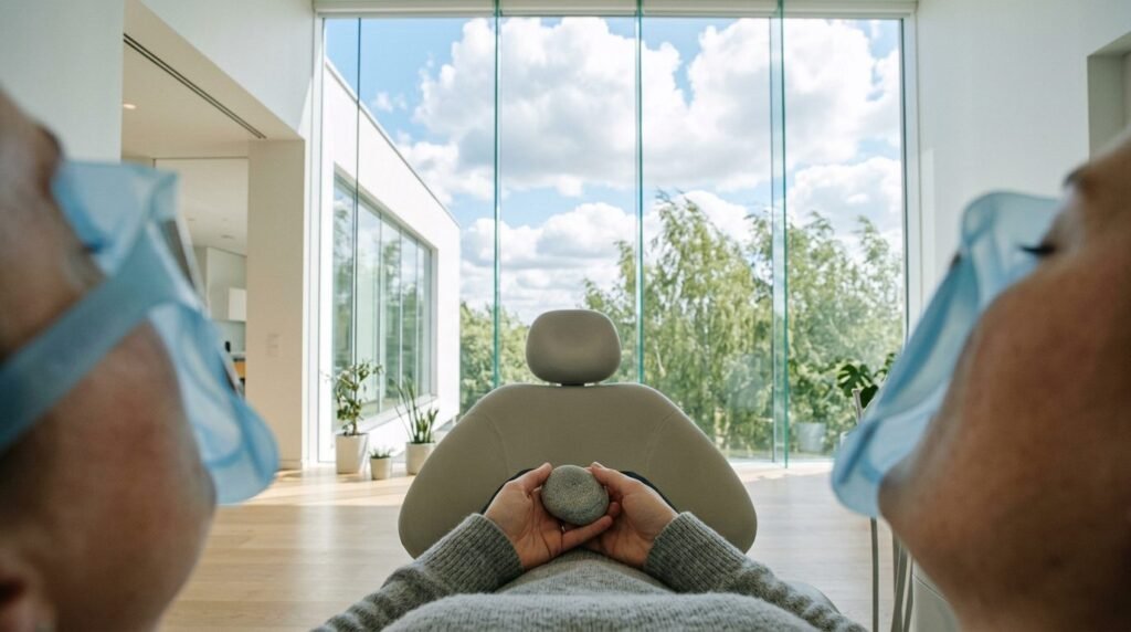
Oral confinement induced by dental isolation equipment triggers acute autonomic claustrophobia by simulating upper airway obstruction. When practitioners stretch rubber dam sheets across metal frames or pack oral vestibules with dense cotton rolls, patients susceptible to spatial confinement experience immediate existential panic. Dismissing patient panic as uncooperativeness overlooks that sealing the oral cavity removes the primary fallback for ventilation while restricting lingual mobility and deglutition. Humanized dentistry recognizes that oral confinement panic represents an innate suffocation alarm mediated by brainstem chemoreceptors and limbic fear circuits. Managing claustrophobic patients requires neurobiological understanding, graded exposure protocols, and modified moisture-control technologies that preserve airway autonomy.
Neurobiology of Oral Confinement: The Suffocation Alarm and Airway Threat
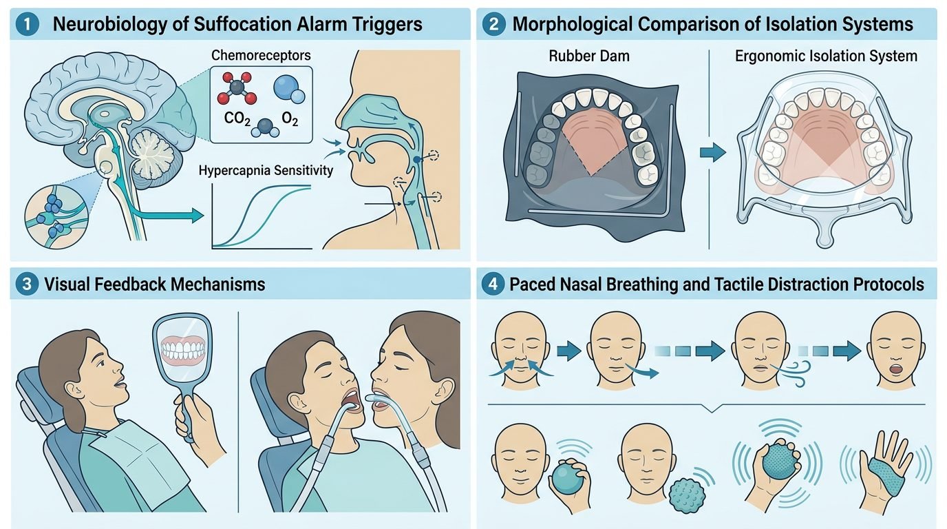
The human brain monitors airway patency through interoceptive networks coordinated by the nucleus of the solitary tract, parabrachial nucleus, and basolateral amygdala. When oral access is abruptly sealed while nasal passages remain partially congested by allergic rhinitis, septal deviation, or supine mucosal engorgement, arterial carbon dioxide tension rises. Brainstem chemoreceptors detect micro-elevations in hypercapnia, discharging rapid warning bursts to higher limbic centers.
This firing triggers the suffocation false alarm mechanism. The amygdala activates massive sympathetic discharge through the locus coeruleus, releasing norepinephrine across the neuroaxis. Within seconds, the patient experiences overwhelming tachycardia, cold diaphoresis, hyperventilation, and an urge to tear the isolation apparatus from the face. Attempting to force compliance by verbally commanding a hypercapnic, panic-stricken patient to relax demonstrates fundamental clinical ignorance. The subjective terror of impending asphyxiation overrides neocortical reasoning. The reaction is an involuntary physiological survival reflex.
Mechanical and Sensory Triggers: Rubber Dams, Cotton Rolls, and Clamps
Specific isolation hardware creates distinct sensory inputs that aggravate confinement phobias. Traditional dark latex rubber dams stretched over rigid Young frames obscure peripheral vision, occlude anterior lighting, and cover the labial commissures completely. The inability to communicate verbally intensifies feelings of helplessness.
Salivary pooling behind the rubber barrier presents an immediate physical threat. When the rubber dam inverts along lingual gingival margins, accumulated saliva and irrigation water pool against the posterior pharyngeal wall. Touching the uvula triggers violent coughing and gagging reflexes. Cotton rolls packed into buccal and lingual vestibules absorb saliva rapidly, adhering tenaciously to dry non-keratinized alveolar mucosa. When clinicians adjust these rolls without rehydration, mechanical shearing of mucosal epithelium causes sharp pain that magnifies sensory hypervigilance. Suction tips wedged into retromolar areas generate localized mucosal suction trauma, creating sensations of physical choking.
Autonomic Deconditioning: Graded Sensory Desensitization Protocols
Overcoming oral confinement anxiety requires systematic autonomic deconditioning using graduated behavioral exposure. Desensitization should be conducted across dedicated familiarization visits rather than attempted during urgent restorative appointments. The protocol begins by deconstructing the rubber dam apparatus into separate physical components, allowing the patient to handle each piece before intraoral placement.
The clinician introduces the rubber dam sheet without the metal frame, holding the perforated elastomeric sheet lightly against a single anterior tooth for ten seconds while the patient breathes through the nose. Once autonomic stability is confirmed by normal respiration, the clamp is seated gently without local anesthesia to demonstrate that placement is painless. In the subsequent phase, the clinician utilizes a clear silicone dam sheet mounted on a collapsible frame. The clear material permits full binocular vision, eliminating dark sensory enclosure. A breathing hole measuring two centimeters in diameter is punched into the contralateral corner of the dam, guaranteeing an immediate open oral airway for emergency mouth breathing. Proving to the patient that airflow remains unhindered disarms the brainstem suffocation reflex.
Modified Isolation Strategies: Balancing Adhesive Quality and Patient Autonomy
Modern clinical restorative dentistry offers versatile moisture-control technologies that maintain high adhesive bonding standards without requiring complete physical confinement. When full rubber dam isolation proves physiologically intolerable despite graded exposure, practitioners must deploy validated alternative isolation devices.
High-volume evacuation isolation mouthpieces made of soft medical-grade silicone integrate tongue retraction, cheek protection, and continuous bilateral suction into a single open-architecture device. These mouthpieces illuminate the oral cavity while maintaining a wide, unobstructed contralateral airway, allowing patients to breathe through mouth and nose simultaneously. For cervical restorations, retraction cords soaked in aluminum chloride paired with localized matrix systems and parotid absorbent shields eliminate rotary contamination without obstructing the dental arch. Split-dam techniques isolating only two adjacent teeth with exposed labial borders provide sufficient moisture control for composite curing while leaving the oral floor completely free for voluntary swallowing.
Communication Strategies and Real-Time Interoceptive Control
Clinical dialogue with confinement-sensitive patients must emphasize constant transparency and absolute physical control. The clinician establishes a bilateral agreement before applying isolation: the patient holds the suction handpiece or an emergency signal bell throughout the procedure. If discomfort arises, the patient activates the device, and the dental team halts instrumentation within two seconds.
Practitioners must maintain verbal contact every sixty seconds, narrating procedure duration: “We are curing the resin for twenty seconds; you have fifteen seconds remaining.” Time estimation becomes distorted during sympathetic arousal; providing external time landmarks prevents panic escalation. The patient is coached in diaphragmatic breathing: inhaling through the nose for four seconds and exhaling slowly through the nose for six seconds. Prolonged exhalation activates the ventral vagal brake, reducing heart rate and damping amygdalar hyperarousal.
Interprofessional Consideration: Respiratory and Structural Airway Screening
Claustrophobic reactions in the dental operatory frequently mask underlying airway pathology. Patients reporting intense dental confinement anxiety exhibit high rates of undiagnosed obstructive sleep apnea, chronic allergic rhinosinusitis, and hypertrophic inferior turbinates. When such patients recline supine, gravity pulls the soft palate and tongue base backward, narrowing pharyngeal space.
Pre-treatment screening should incorporate the STOP-Bang questionnaire and a physical examination of the oropharynx using the Mallampati classification. When anatomical airway obstruction is recognized, the clinician plans operative sessions in a semi-upright chair angle of forty-five degrees and encourages pre-operative nasal decongestant sprays. Coordinating care with an otolaryngologist addresses the underlying structural breathing deficit, transforming dental isolation from a terrifying ordeal into a routine, comfortable procedure.
Clinical Synthesis for the Contemporary Dental Team
Humanized dentistry rejects the false dichotomy between uncompromising technical excellence and patient emotional safety. Preserving the bond strength of modern resin composites does not require inflicting psychic trauma or ignoring physiological panic. By understanding the neurobiology of suffocation fear, modifying physical isolation equipment, and sharing procedural control with the patient, the dental team delivers restorative dentistry that protects oral tissues while honoring human dignity.
References
- Hill, F. J., & O’Mullane, D. M. (1976). The use of rubber dam in pediatric dentistry. British Dental Journal, 141(9), 273-278.
- Klein, D. F. (1993). False suffocation alarms, spontaneous panics, and related conditions: an integrative hypothesis. Archives of General Psychiatry, 50(4), 306-317.
- Pini, N. I., et al. (2014). Rubber dam isolation in restorative dentistry: clinical advantages and patient comfort. General Dentistry, 62(5), 36-41.
- De Jongh, A., et al. (2002). Psychological management of the claustrophobic dental patient: a case study. Journal of Anxiety Disorders, 16(4), 455-463.
- Anusavice, K. J., et al. (2013). Phillips’ Science of Dental Materials. Elsevier Health Sciences.
Leave a Reply