
Carbon dioxide (CO2) laser surgery provides superior hemostasis, reduced post-operative edema, and minimal myofibroblastic scarring compared to cold-blade scalpel excision when treating potentially malignant oral disorders, specifically oral leukoplakia. However, deploying infrared laser energy across dysplastic mucosa introduces diagnostic dilemmas and technical compromises that clinicians must critically manage. Vaporization eliminates the lesion entirely through photothermal dehydration and carbonization, but deprives the oral pathologist of a tissue specimen for histological staging, creating risks of missing occult microinvasive squamous cell carcinoma. Conversely, excisional biopsy performed with CO2 lasers introduces thermal coagulation artifacts along surgical margins, potentially obscuring cellular dysplasia and basilar membrane architecture. Furthermore, eliminating visible keratotic plaques with laser ablation does not eliminate underlying genetic field cancerization within clinically normal adjacent mucosa. Long-term disease control depends on understanding the optical absorption physics of 9300 nm versus 10,600 nm wavelengths, establishing standardized ablation depths, and enforcing strict longitudinal surveillance protocols.
The biophysical distinction between 9300 nm and 10,600 nm carbon dioxide lasers resides in their optical absorption coefficients in oral soft tissue chromophores. The classical 10,600 nm wavelength corresponds to an absorption coefficient in pure water of approximately eight hundred inverse centimeters, yielding an optical penetration depth of roughly twelve to fifteen micrometers. In contrast, the 9300 nm isotopic CO2 wavelength coincides precisely with the fundamental infrared absorption band of both water and the phosphate groups within hydroxyapatite, exhibiting an absorption coefficient exceeding five thousand inverse centimeters. This massive absorption coefficient restricts photonic energy penetration to fewer than three to five micrometers. Clinically, the 9300 nm beam produces rapid tissue vaporization with a significantly narrower lateral thermal collateral damage zone (twenty to fifty micrometers) compared to the 10,600 nm beam (one hundred to two hundred micrometers). For excisional biopsies, the narrower thermal margin of the 9300 nm laser preserves fine cytological detail at the specimen perimeter, allowing pathologists to evaluate cellular pleomorphism and mitotic figures right up to the surgical cut without confounding coagulation artifacts.
Clinical management mandates a clear diagnostic sequence: incisional biopsy must always precede extensive laser vaporization. In homogeneous leukoplakias exhibiting uniform, thin, flat hyperkeratosis, a representative cold scalpel or punch biopsy confirms the presence or absence of epithelial dysplasia. When mild or no dysplasia is identified, superficial vaporization of the entire lesion represents a valid, tissue-preserving treatment strategy. However, non-homogeneous leukoplakias characterized by erythroleukoplakic mottling, verrucous elevations, or nodular ulcerations exhibit malignancy transformation rates up to five times higher than homogeneous variants. In non-homogeneous presentations, blind vaporization is contraindicated. The clinician must execute a complete excisional biopsy with an intact peripheral clinical safety margin of at least three to five millimeters of healthy tissue, extending down into the underlying submucosa or muscular layer to ensure comprehensive histological assessment.
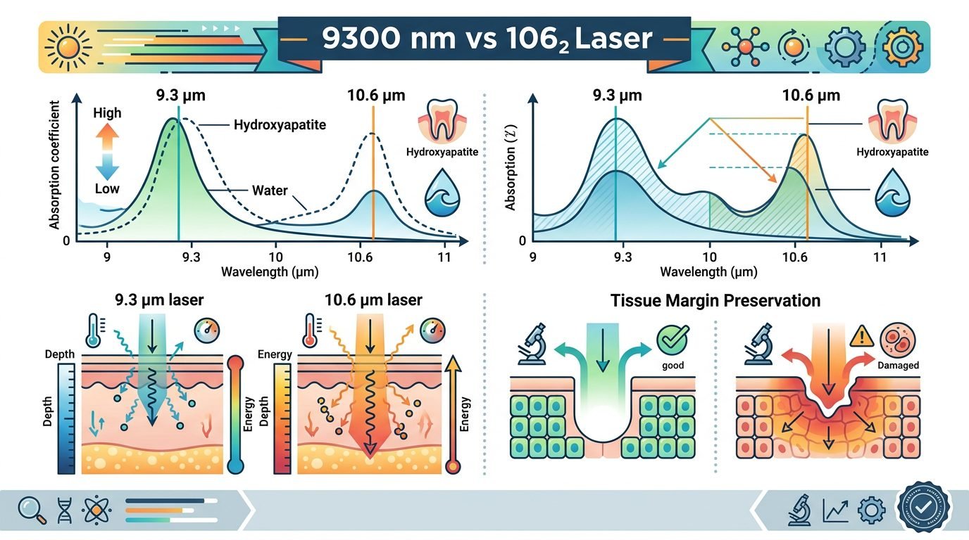
Ablation depth directly dictates both surgical radicality and functional recurrence rates. Epithelial dysplasia frequently extends into the epithelial rete pegs and along minor salivary gland excretory ducts. Vaporization restricted to the superficial stratum corneum or prickle cell layer leaves genetically altered basal progenitor cells intact, guaranteeing rapid lesion recurrence. Clinical studies utilizing intraoperative optical coherence tomography (OCT) or frozen-section biopsy demonstrate that therapeutic laser ablation must penetrate into the upper papillary and reticular lamina propria, achieving a uniform surgical crater depth between 1.0 and 1.5 millimeters across mucosal surfaces, and up to 2.0 millimeters on the thick ventral tongue or buccal mucosa. Ablating beyond 2.0 millimeters in non-malignant dysplasia provides no additional oncological security while dramatically increasing the risk of collateral damage to underlying lingual motor nerves, branches of the facial artery, and deep salivary ducts, which leads to sialocele formation or permanent sensory paresthesias.
Recurrence rates following CO2 laser treatment highlight the biological realities of mucosal field cancerization. Systematic reviews and meta-analyses encompassing thousands of treated leukoplakia cases reveal recurrence rates ranging between twelve and twenty-nine percent following complete laser excision or vaporization, with malignant transformation occurring in three to seven percent of cases over a five-year follow-up interval. These recurrence rates reflect the persistent presence of clonal genetic alterations, including mutations in the TP53 gene and chromosomal loss of heterozygosity (LOH) at loci 9p21 and 3p14, residing within histologically normal-appearing mucosal margins beyond the visible surgical crater. Clinicians who interpret laser wound clearance as a definitive cancer cure misunderstand cancer biology. Laser ablation eliminates the high-risk macroscopic lesion with minimal morbidity, but the patient remains systemically predisposed to secondary primary lesions across the entire upper aerodigestive tract.
Operational parameters must balance cutting velocity against peripheral thermal coagulation. When utilizing a 10,600 nm CO2 laser for excisional biopsy, focused micro-spot sizes between 0.2 and 0.4 millimeters delivering continuous or super-pulsed average powers of five to eight watts produce rapid incisions with clean hemostatic sealing of capillaries and lymphatic vessels smaller than 0.5 millimeters. For broad-field vaporization of low-risk homogeneous plaques, a defocused beam spot size of 1.0 to 2.0 millimeters operating at four to six watts in a continuous sweeping cross-hatch pattern ensures controlled layer-by-layer sublimation. Continuous high-volume intraoral evacuation equipped with specialized sub-micron viral and smoke particulate filters (ULPA filtration) is mandatory to capture toxic surgical plumes containing volatile organic compounds, viable cellular DNA fragments, and hazardous nanoparticles.
Post-operative healing characteristics distinguish CO2 laser surgery from conventional cold scalpel techniques. The surgical crater heals by secondary intention without primary suturing. Because the CO2 laser seals peripheral nerve endings and creates a superficial coagulated proteinaceous coagulum, post-operative pain scores are remarkably lower than those following scalpel excision. By day three post-surgery, a thick, yellow-white fibrinous pseudomembrane covers the ablated zone, which inexperienced clinicians often mistake for acute bacterial infection or recurrent candidiasis; this fibrinous matrix represents normal secondary granulation and must not be debrided. Re-epithelialization proceeds via centripetal migration from wound margins, completing within fourteen to twenty-one days with remarkably supple, uncontracted mucosal architecture. Furthermore, the absence of deep myofibroblastic scar bands prevents functional tongue restriction or severe trismus when treating lesions adjacent to the pterygomandibular raphe or retro-molar trigone. Patients maintain normal speech, swallowing dynamics, and oral opening throughout the convalescent period.
Regarding comprehensive clinical protocols, carbon dioxide lasers operating at 9300 nm and 10,600 nm represent standard surgical instruments for managing oral leukoplakia. Their physical superiority in bloodless cutting and low-scarring healing is unmatched. Nevertheless, laser technology cannot compensate for flawed diagnostic judgment. Clinicians must maintain a strict biopsy-first protocol for high-risk lesions, respect anatomical ablation depths, ensure unburned margins during excisional procedures, and enforce indefinite longitudinal recalls every three to six months to intercept recurrent dysplasia and field malignant transformations. Consistent clinical surveillance combined with targeted lifestyle modifications, including complete smoking cessation and alcohol abstinence, remains the true foundation of long-term oncological prevention in oral mucosal pathology.
Leave a Reply