

Providing dental care to patients with end-stage renal disease undergoing maintenance hemodialysis requires precise scheduling around systemic heparinization, strict protection of arteriovenous vascular access, and tailored pharmacological dosing. Chronic kidney disease (CKD) stages 4 and 5 fundamentally alters systemic homeostasis, creating profound hematological, cardiovascular, immunological, and metabolic dysfunctions. Individuals on hemodialysis face elevated bleeding risks from uremic platelet dysfunction and systemic anticoagulation, alongside severe susceptibility to bacterial infections and secondary hyperparathyroidism. In the dental operatory, procedural errors such as taking blood pressure on an access arm, scheduling extractions on dialysis days, or prescribing nephrotoxic analgesics can precipitate vascular access thrombosis, refractory hemorrhage, or fatal acute toxicity. Establishing safe clinical outcomes demands integrating nephrological pharmacokinetics with humanized, supportive patient scheduling.
Renal Osteodystrophy and Uremic Stomatological Manifestations
End-stage renal disease generates pronounced hard and soft tissue oral pathologies driven by impaired calcitriol synthesis, phosphate retention, and circulating uremic toxins. Decreased renal activation of vitamin D leads to hypocalcemia, triggering secondary hyperparathyroidism. Excess parathyroid hormone resorbs trabecular bone, manifesting radiographically as loss of the lamina dura, ground-glass trabecular demineralization, and localized radiolucent osteolytic lesions termed brown tumors of hyperparathyroidism.
Soft tissue manifestations reflect uremic toxicity and severe xerostomia. Salivary urea levels rise proportionally to blood urea nitrogen (BUN), where bacterial ureases hydrolyze urea into ammonia. This process generates a characteristic uremic fetor and metallic dysgeusia. Furthermore, strict fluid restriction (often under 500 to 1,000 mL per day) depresses salivary flow, precipitating mucosal atrophy, candidiasis, and painful uremic stomatitis characterized by white, non-wipeable hyperkeratotic mucosal plaques. Clinicians must recognize these oral signs as reflections of severe metabolic strain rather than isolated dental infections.
Anticoagulation Kinetics: The Hemodialysis Window and Heparin Half-Life
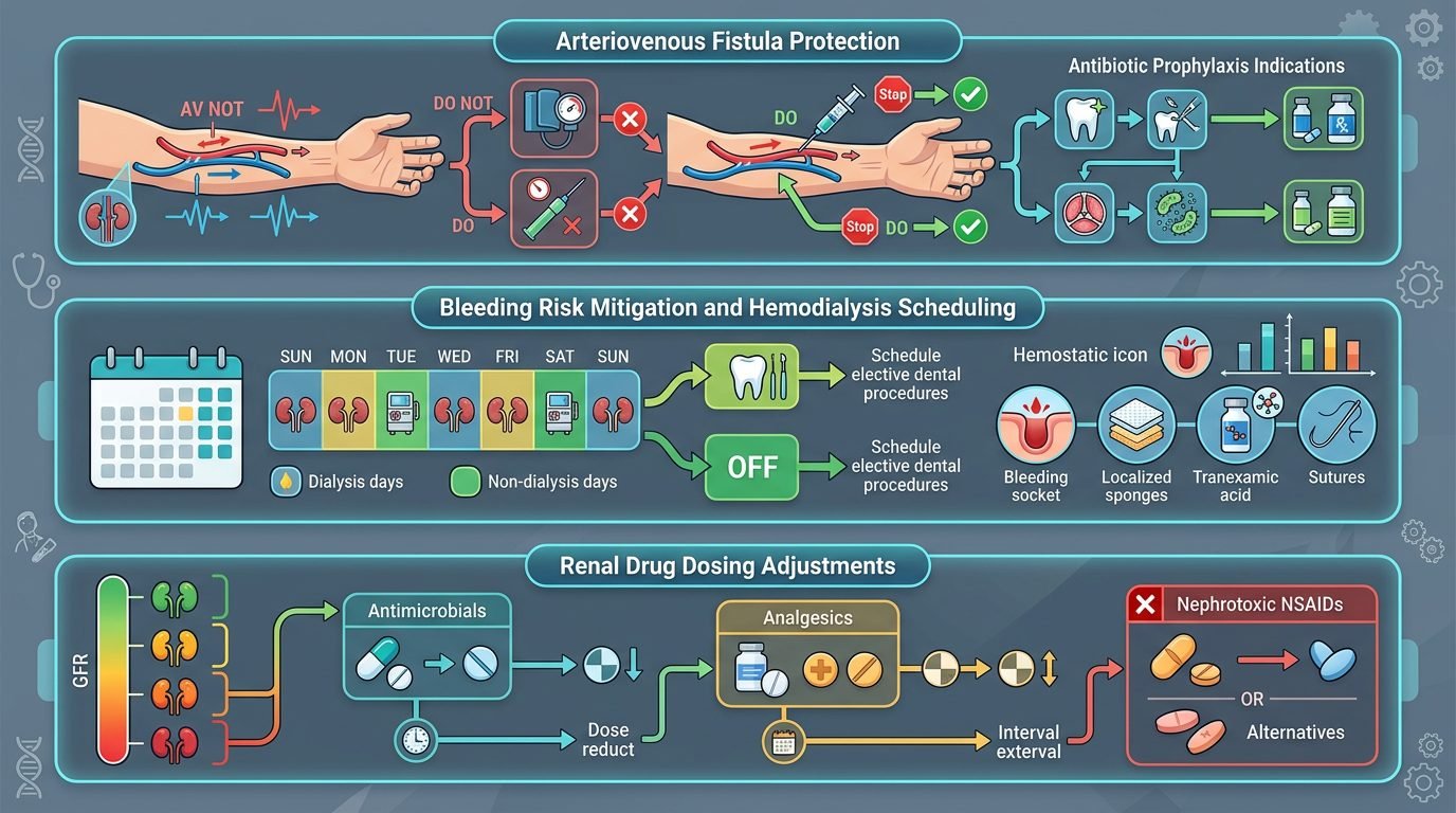
Managing bleeding risk centers on the pharmacokinetics of systemic heparin administered during hemodialysis sessions to prevent extracorporeal circuit clotting. Unfractionated heparin possesses an elimination half-life of 60 to 90 minutes, whereas low-molecular-weight heparins exhibit half-lives of three to six hours. Performing invasive dental procedures on the day of hemodialysis invites catastrophic hemorrhage, as circulating heparin combines with pre-existing uremic thrombocytopathy to paralyze the clotting cascade.
The standard clinical window for invasive dental treatment is the day following hemodialysis (the “off-dialysis” day). For example, if dialysis occurs on Monday, invasive procedures should be performed on Tuesday morning. By this time, exogenous heparin has undergone complete metabolic clearance, while fluid overload, hyperkalemia, and uremic toxin spikes have not yet reached peak pre-dialysis levels. Invasive treatment must never be scheduled immediately prior to a dialysis session, as active clot formation will be lysed when full-dose heparin is infused hours later during dialysis.
Arteriovenous Fistula Preservation and Infection Control
The arteriovenous (AV) fistula or synthetic graft represents the patient’s lifeline for vascular access, demanding absolute surgical preservation. Occlusion, damage, or bacterial infection of an AV fistula requires urgent vascular surgery and precipitates life-threatening interruptions in dialysis therapy.
Dental professionals must enforce strict physical precautions: the arm bearing the AV fistula or graft must never be utilized for blood pressure cuff placement, intravenous catheterization, or venipuncture. Compressive pressure on the fistula can induce immediate intraluminal thrombosis. The patient should not rest their head or body weight upon the access limb in the dental chair. Although routine antibiotic prophylaxis for dental procedures in patients with native AV fistulas is no longer recommended by the American Heart Association unless synthetic polytetrafluoroethylene (PTFE) grafts were placed within the previous six months, the patient’s nephrologist should be consulted regarding individualized bacteremia prevention protocols, particularly in the presence of active odontogenic infections.
Pharmacological Dosing Adjustments in Renal Failure
Prescribing medications in patients with severely reduced glomerular filtration rates (eGFR under 15 mL/min/1.73m2) requires rigorous dosage modification. Nephrotoxic nonsteroidal anti-inflammatory drugs (NSAIDs), including ibuprofen, naproxen, and ketorolac, are strictly contraindicated; NSAIDs further compromise residual nephron perfusion, trigger gastrointestinal bleeding in uremic mucosa, and blunt antihypertensive therapy. Tetracyclines (with the exception of doxycycline) should also be avoided because their anti-anabolic effect accelerates amino acid catabolism, sharply increasing blood urea nitrogen levels and worsening uremic symptoms.
Acute odontogenic pain must be managed with acetaminophen, maintaining total daily doses below two grams to prevent hepatotoxicity, or short courses of tramadol adjusted for renal clearance. For odontogenic infections, amoxicillin and cephalexin remain first-line choices but require extended dosing intervals (every 12 to 24 hours instead of every 8 hours) to prevent neurotoxicity and crystal accumulation. Clindamycin, azithromycin, and metronidazole undergo predominantly hepatic metabolism and do not require renal dose adjustments, serving as reliable alternatives.
Hemostatic Protocols for In-Office Extractions
Even on off-dialysis days, qualitative platelet dysfunction (caused by abnormal platelet-endothelial adhesion and impaired von Willebrand factor binding) predisposes to persistent post-extraction oozing. Relying solely on simple gauze compression is inadequate.
Clinicians must employ aggressive local hemostatic measures: surgical sockets must be packed with oxidized regenerated cellulose, resorbable gelatin sponges impregnated with topical thrombin, or microfibrillar collagen. Multiple interrupted horizontal mattress sutures should be placed under slight tension to stabilize mucosal flaps. In cases of severe refractory uremic oozing, coordinating with the nephrologist for an intravenous or intranasal dose of desmopressin (DDAVP) stimulates the release of endothelial von Willebrand factor and factor VIII, transiently normalizing primary hemostasis. Furthermore, the patient should rinse with 4.8% tranexamic acid mouthwash four times daily for five days without swallowing the solution. This antifibrinolytic barrier inhibits salivary plasminogen activation, preventing clot dissolution during post-extraction recovery.
Empathetic Scheduling and Dialysis Fatigue
Beyond surgical technicalities, humane care requires recognizing the extreme physical exhaustion (“dialysis fatigue”) that follows a four-hour filtration session. Patients endure fluid shifts, hypotensive episodes, muscle cramping, and mental fatigue that leave them bedridden for hours post-treatment.
The dental team must demonstrate operational flexibility, avoiding rigid cancellation penalties when unexpected access revisions or medical emergencies arise. Appointments should be kept brief (30 to 45 minutes) and scheduled during the patient’s peak energy window, typically mid-morning on non-dialysis days. By accommodating the rigorous rhythm of hemodialysis with technical expertise and deep biological empathy, the dental team protects vital vascular lines while restoring health and dignity to medically complex patients.
References
- De Rossi, S. S., & Glick, M. (1996). Dental considerations for the patient with renal disease receiving hemodialysis. Journal of the American Dental Association, 127(2), 211-219.
- Kerr, A. R. (2001). Update on renal disease for the dental practitioner. Oral Surgery, Oral Medicine, Oral Pathology, Oral Radiology, and Endodontics, 92(1), 9-16.
- Little, J. W., et al. (2018). Dental Management of the Medically Compromised Patient (9th ed.). Elsevier.
- Proctor, R., et al. (2005). Oral manifestations of gastrointestinal and renal diseases. Periodontology 2000, 39(1), 40-52.
- Thomas, C. M., & Fabbroni, G. (2020). The dental management of patients with chronic kidney disease. Dental Update, 47(4), 312-319.
Leave a Reply