
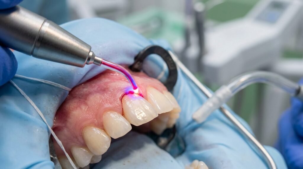
Surgical gingival recontouring in the maxillary esthetic zone requires precise architectural control and immediate vascular stabilization. Patients presenting with excessive gingival display, altered passive eruption, or coronal asymmetry demand harmonious parabolic margins without postoperative recession, prolonged bleeding, or open wound complications. Traditional cold-blade scalpels produce profuse microvascular hemorrhage that obscures the operative field, precludes immediate adhesive bonding, and necessitates non-esthetic periodontal dressings. Electrosurge units provide hemostasis but introduce severe lateral heat conduction exceeding one hundred degrees Celsius, risking periodontal ligament necrosis, alveolar bone resorption, and unpredictable gingival margin slump. Semiconductor diode lasers, operating across near-infrared wavelengths (810, 940, and 980 nm) and the visible blue spectrum (445 nm), deliver precise photothermal soft tissue vaporization. This biophysical mechanism seals capillary loops instantaneously, preserves supracrestal tissue attachment, and accelerates secondary intention re-epithelialization with minimal postoperative patient discomfort.
Photobiophysics of Diode Laser Cutting and Chromophore Absorption
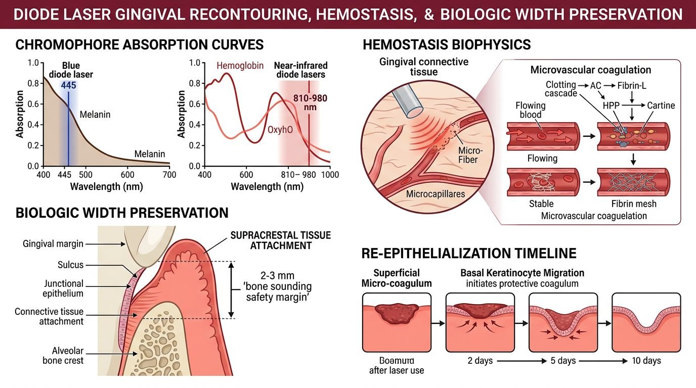
Semiconductor diode lasers deliver coherent monochromatic optical radiation through flexible quartz optical fibers ranging from two hundred to four hundred micrometers in diameter. In biological oral tissues, diode photon absorption is governed by primary tissue chromophores, specifically oxyhemoglobin, deoxyhemoglobin, and melanin. Near-infrared emissions between 810 and 980 nanometers exhibit low optical absorption in pure water but high affinity for pigmented chromophores in vascularized gingival tissues.
To cut non-pigmented fibrous gingiva efficiently, near-infrared fiber tips must undergo deliberate optical initiation, typically utilizing articulating paper or dedicated carbon blocks. Initiation concentrates thermal energy at the extreme distal glass boundary, converting optical radiation into intense contact thermal cutting. Conversely, visible blue diode lasers operating at 445 nanometers exhibit an absorption coefficient in hemoglobin that is several hundred times higher than near-infrared wavelengths. Blue laser light cuts biological tissues via direct photon-chromophore absorption, allowing instantaneous, non-contact or contact ablation without requiring artificial tip initiation, thereby minimizing superficial tissue charring.
Hemostasis Mechanisms and Immediate Operative Dryness
A primary clinical advantage of diode laser soft tissue sculpting is instantaneous, sustained hemostasis. When the energized fiber incises vascularized papillary and marginal gingival tissue, localized photothermal temperatures reach between one hundred and one hundred and fifty degrees Celsius. This rapid thermal energy delivery denatures soluble plasma fibrinogen, converting it instantly into an insoluble fibrin mesh that seals severed capillaries and venules up to 0.5 millimeters in diameter.
Simultaneously, thermal contraction of perivascular smooth muscle cells collapses lumen diameters, terminating microvascular blood flow immediately. The resulting operative wound bed is entirely dry, clean, and free of blood contamination. This pristine environment allows clinicians to perform immediate digital optical scanning, rubber dam placement, or adhesive resin bonding for porcelain laminate veneers in a single clinical appointment. Eliminating hemorrhage avoids blood staining of resin margins and prevents hydrolytic degradation of adhesive hybrid layers during resin curing.
Biologic Width Preservation and Bone Sounding Protocols
Predictable long-term stability of the recontoured gingival margin requires strict adherence to biological width, anatomically defined as the supracrestal tissue attachment. Histological architecture dictates that the junctional epithelium (averaging 0.97 millimeters) and supracrestal connective tissue attachment (averaging 1.07 millimeters) require a minimum physiological dimension of approximately two millimeters between the alveolar bone crest and the apical base of the gingival sulcus.
Prior to laser ablation, the clinician must execute transgingival bone sounding under local anesthesia, inserting a calibrated periodontal probe through the sulcus to contact the facial bone crest. If bone sounding reveals a supracrestal soft tissue dimension of three millimeters or greater, gingivoplasty can be performed safely with the diode laser without violating the junctional attachment. However, if bone sounding demonstrates less than three millimeters of soft tissue above the crest, closed laser gingivoplasty is contraindicated; performing soft tissue resection in this situation violates the biological width, triggering chronic inflammatory breakdown, perpetual gingival bleeding, or unpredictable marginal bone resorption. In such clinical presentations, a full-thickness flap with osseous recontouring remains mandatory.
Cellular Wound Healing Dynamics and Thermal Damage Zones
The biological quality of mucosal wound healing following diode laser surgery depends entirely on managing the zone of collateral thermal damage. Histological analyses show that a properly calibrated diode laser creates an ultra-thin layer of superficial coagulative necrosis measuring between fifty and one hundred micrometers in thickness. This micro-carbonized coagulum acts as a biological dressing that shields underlying exposed nerve endings from oral fluids, explaining why patients report virtually zero postoperative pain.
Within forty-eight hours post-surgery, neutrophils clear the minimal necrotic zone while keratinocytes migrate beneath the coagulum. By seven to ten days, a fully stratified, parakeratinized oral epithelium covers the surgical site. Transforming growth factor-beta and basic fibroblast growth factor expression remain optimized, preventing the excessive myofibroblastic differentiation responsible for surgical scar contracture. The healed tissue exhibits natural stippling, harmonious color matching, and stable scalloped gingival contours identical to healthy native gingiva.
Operatory Application Protocols and Fiber Management
Achieving optimal tissue sculpting without excessive thermal necrosis demands refined clinical technique. For near-infrared diode systems (810 to 980 nm), the clinician operates in pulsed or continuous-wave mode with average power settings kept strictly between 0.8 and 1.2 watts. The initiated fiber tip should be held at a forty-five-degree angle relative to the long axis of the tooth, moving in rapid, light feather-like brush strokes across the tissue margin.
The operator must never hold the energized fiber stationary against the gingiva, as heat accumulation induces deep lateral carbonization and delayed tissue healing. High-volume evacuation should be positioned within two centimeters of the ablation site to remove the bio-aerosol laser plume efficiently. Regular cleaning of the fiber tip with a moist sterile gauze removes carbonized tissue debris, maintaining sharp optical focus and preventing excessive thermal accumulation. Postoperatively, patients require only mild saline rinses and gentle mechanical brushing, avoiding the need for periodontal dressings or systemic analgesics.
Clinical Considerations in Restorative Smile Design
Diode laser gingivoplasty bridges the boundary between periodontal surgery and restorative smile design. By delivering bloodless soft tissue ablation with sub-millimeter precision, the clinician eliminates procedural bleeding, protects the supracrestal biological attachment, and ensures rapid re-epithelialization. Deploying calibrated diode energy establishes a stable, biologically sound foundation for enduring esthetic outcomes.
References
- Goharkhay, K., et al. (1999). Soft tissue surgery in the oral cavity with a diode laser: an in vivo study. Lasers in Medical Science, 14(4), 263-269.
- Romanos, G. E., & Nentwig, G. H. (1999). Diode laser (980 nm) in oral and maxillofacial surgical procedures: clinical observations based on clinical applications. Journal of Clinical Laser Medicine & Surgery, 17(5), 193-197.
- Garg, K., et al. (2014). Diode laser in management of esthetic gingival problems: a case series. Journal of Dental Lasers, 8(1), 35-39.
- Amaral, M. B., et al. (2015). Diode laser surgery in the management of oral lesions: a clinical study. Journal of Cranio-Maxillofacial Surgery, 43(8), 1621-1627.
- Fornaini, C., et al. (2016). 445 nm diode laser in oral surgery: a preliminary in vitro study. Photomedicine and Laser Surgery, 34(11), 548-554.
Leave a Reply