
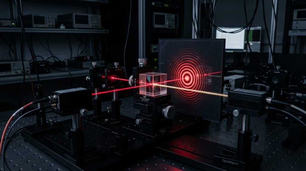
Bilateral sagittal split ramus osteotomy (BSSO) represents the primary surgical intervention for severe skeletal maxillomandibular discrepancies, yet persistent neurosensory disturbance of the inferior alveolar nerve (IAN) remains its most frequent complication. Mechanical manipulation during osteotomy splitting, nerve traction, and direct compression from bicortical screws induce neurapraxia or axonotmesis in up to eighty-five percent of patients immediately post-surgery. Prolonged hypoesthesia of the lower lip and chin impairs oral competence, mastication, and psychosocial quality of life for months or years. Adjunctive dual-wavelength photobiomodulation (PBM) combining visible red (660 nm) and near-infrared (808 nm) radiation provides a biophysical method to accelerate axonal regeneration, reduce postoperative inflammatory edema, and promote osteogenesis along the mandibular osteotomy gap.
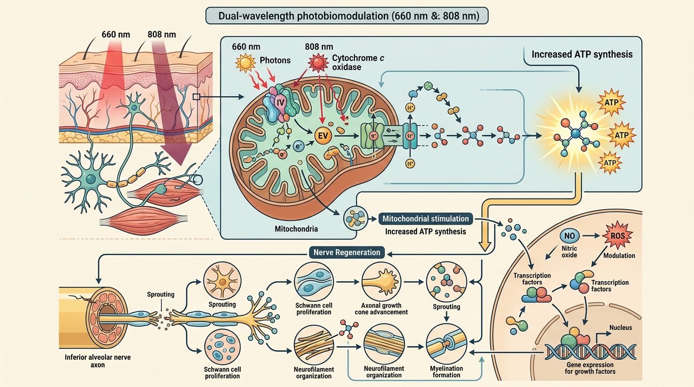
The biological rationale for combining 660 nm and 808 nm wavelengths centers on optical penetration depths and differential chromophore absorption in traumatized facial tissues. Photons emitted at the visible 660 nm wavelength are absorbed predominantly in superficial capillary micro-networks, erythrocyte porphyrins, and submucosal connective tissue layers, stimulating local microcirculation and active lymphatic endothelial drainage to resolve acute facial swelling. Conversely, the near-infrared 808 nm wavelength exhibits minimal optical absorption by oxyhemoglobin and water, creating an optical transmission window that facilitates deep photonic penetration through the masseter muscle bundle and dense buccal cortical bone to reach the mandibular canal. At the fundamental mitochondrial level, red and near-infrared photons are absorbed by specific metal centers within unit IV of the respiratory chain, cytochrome c oxidase, specifically the oxidized copper centers (CuA) at 660 nm and the reduced iron-heme centers (Fe-a3) at 808 nm within Schwann cells and neuronal axoplasm. This photon absorption accelerates electron transport chain turnover, elevates adenosine triphosphate (ATP) synthesis, modulates reactive oxygen species (ROS) to physiological signaling levels, and stimulates intracellular cyclic adenosine monophosphate (cAMP) transcription, preventing secondary apoptotic neuronal demise.
Axonal regeneration following mechanical nerve crush depends directly on early Schwann cell proliferation and neurotrophin upregulation. Within the injured inferior alveolar nerve, ischemia and inflammatory cytokine storms trigger Wallerian degeneration distal to the osteotomy site. Experimental and human trials indicate that 808 nm irradiation upregulates the synthesis of nerve growth factor (NGF), brain-derived neurotrophic factor (BDNF), and neurotrophin-3 (NT-3). Concurrently, photobiomodulation accelerates Schwann cell migration along degenerating basal lamina tubes, forming organized bands of Bungner that guide regenerating axonal growth cones toward peripheral terminal fields. Electrophysiological evaluations, including sensory nerve action potential (SNAP) amplitudes and trigeminal somatosensory evoked potentials (TSEP), confirm earlier re-innervation and increased conduction velocity in laser-treated patients compared to sham-irradiated controls.
Clinical neurosensory testing protocols confirm objective, quantifiable functional gains across standardized recovery timelines. Patients undergoing rigorous neurosensory evaluations (including two-point discrimination using calibrated digital Boley calipers, static light touch thresholds assessed with Semmes-Weinstein monofilaments, brush directional tactile discrimination, and thermal discrimination using cold and warm thermal probes) exhibit significantly faster recovery trajectories under active dual-wavelength photobiomodulation therapy. Millimetric calibration in two-point discrimination reveals that while unassisted surgical controls often maintain abnormal spatial thresholds exceeding ten to fifteen millimeters beyond six months, laser-treated cohorts regain normal discriminatory tactile thresholds (under five to seven millimeters) within eight to twelve weeks. By the twelfth postoperative week, over eighty-two percent of laser-treated surgical sites display normal light touch thresholds across the mental and inferior labial dermatomes, compared to forty-six percent in untreated surgical controls. The rapid normalization of sensory inputs mitigates accidental lip-biting trauma, facilitates clear articulation, and restores natural masticatory proprioception during complex bolus chewing.
Facial edema and acute postoperative trismus represent secondary therapeutic targets addressed through combined red and infrared radiation. In the immediate seventy-two hours following orthognathic intervention, intense surgical dissection triggers vascular hyperpermeability and interstitial fluid accumulation. Red laser irradiation at 660 nm enhances endothelial nitric oxide synthase (eNOS) activity, facilitating localized vasodilation followed by enhanced lymphatic endothelial contractility that clears interstitial macromolecular exudates. Three-dimensional stereophotogrammetry demonstrates a thirty to forty-two percent reduction in facial soft-tissue volumetric swelling on postoperative days three and seven in patients receiving protocol-driven PBM. Concurrently, accelerated resolution of masseteric inflammation allows faster return of inter-incisal mouth opening.
Osteogenic stimulation across the mandibular osteotomy gap represents an equally vital clinical advantage of near-infrared irradiation. Stable osseous consolidation between proximal and distal mandibular segments is necessary to avoid skeletal relapse and ensure long-term occlusal stability. Photons penetrating to the bony osteotomy line stimulate osteoblastic differentiation from local mesenchymal stem cells. This photonic stimulation enhances alkaline phosphatase enzymatic expression, elevates osteocalcin synthesis, and upregulates bone morphogenetic proteins (BMP-2 and BMP-7). Radiographic density analyses utilizing serial cone-beam computed tomography (CBCT) demonstrate enhanced bone bridging and earlier cortical remineralization along the sagittal osteotomy interface at eight and sixteen weeks post-surgery.
Dosimetric parameters must be accurately calibrated to ensure adequate target fluence without causing thermal accumulation or inhibitory tissue saturation. Clinical delivery requires a multi-point transcutaneous and intraoral irradiation protocol initiated within twenty-four hours of surgery and continued three times weekly across four consecutive weeks, allowing forty-eight to seventy-two hours of cellular recovery between sessions. Intraoral applications utilize an 808 nm continuous-wave gallium-aluminum-arsenide (GaAlAs) diode laser delivering an irradiance of one hundred milliwatts per square centimeter over three distinct mucosal points along the mandibular ramus osteotomy line, applying an energy density of 4 to 6 joules per square centimeter per target point with a spot size of 0.5 square centimeters. Transcutaneous irradiation applies combined 660 nm and 808 nm cluster probes along the anatomical pathway of the inferior alveolar and mental nerves, targeting the mandibular angle, mid-ramus, and the mental foramen. The total cumulative energy per treatment session must remain between twenty and thirty joules per mandibular quadrant to avoid the biphasic Arndt-Schulz inhibitory response, where excessive photonic dosing dampens cellular bioenergetics.
Safety precautions and clinical contraindications require strict monitoring throughout orthognathic laser rehabilitation. The treating surgical team must verify that rigid internal osteosynthesis fixation hardware, such as titanium miniplates or bicortical screws, does not induce dangerous focal thermal reflection or localized overheating of adjacent soft tissues. Continuous contact scanning or non-contact perpendicular irradiation with wavelength-specific certified safety goggles (optical density greater than 5+ at both 660 nm and 808 nm) is strictly mandatory for the patient, operator, and surgical assistants. Photobiomodulation must be rigorously avoided in patients with active malignant lesions or prior history of head and neck oncological resections directly within the craniofacial irradiation field.
Regarding clinical practice, incorporating dual-wavelength photobiomodulation into standard orthognathic surgical pathways establishes a non-invasive, evidence-based modality to accelerate comprehensive patient recovery. By mitigating persistent inferior alveolar nerve paresthesias, rapidly resolving acute inflammatory facial edema, and accelerating early mandibular osteotomy osseous union, dual-wavelength photonic irradiation significantly improves both objective physiological healing and subjective patient quality of life. Maxillofacial surgical teams should routinely adopt standardized, reproducible dosimetric protocols to transform post-surgical outcomes and elevate neurosensory rehabilitation beyond conventional expectant observation.
Leave a Reply