
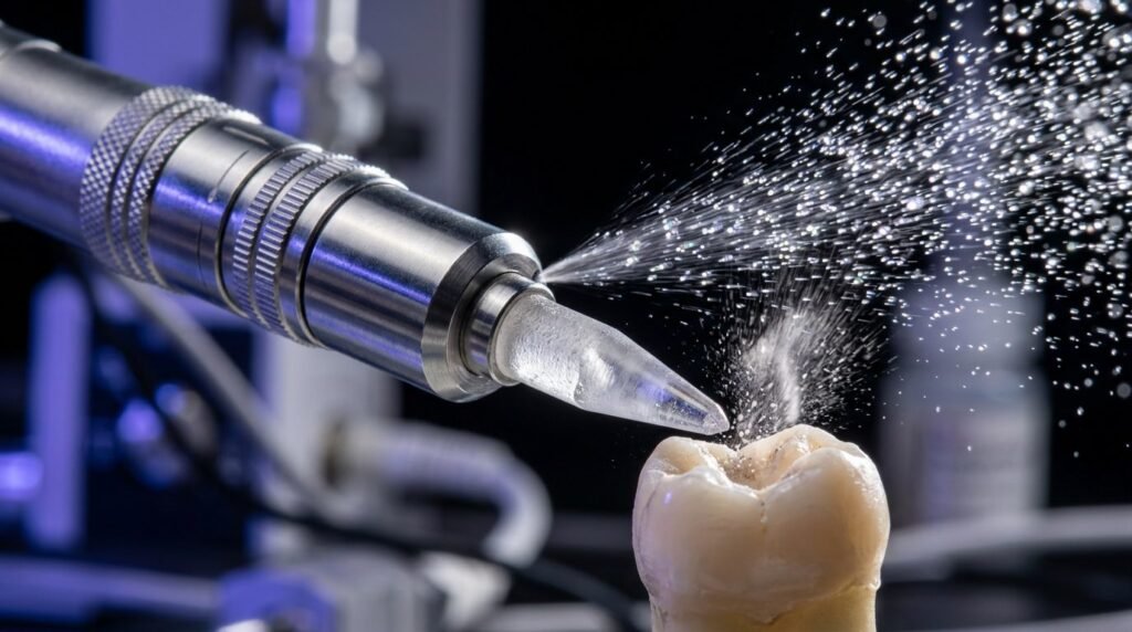
Erbium lasers alter hard dental tissue substrates by thermomechanical micro-explosions rather than chemical demineralization, producing distinct micro-retentive surface morphologies. The interaction of Er:YAG (2,940 nm) and Er,Cr:YSGG (2,780 nm) wavelengths with biological hydroxyapatite and interstitial water challenges standard adhesive protocols established for phosphoric acid etching. While phosphoric acid dissolves mineral to expose vulnerable collagen, erbium ablation eliminates the smear layer and opens tubules cleanly without demineralization. However, laser preparation produces subsurface microcracking and altered protein structures that compromise initial bond strength if clinicians fail to adjust pulse duration and water irrigation. Evaluating the micro-morphological reality of laser-conditioned enamel and dentin is necessary for achieving predictable long-term clinical adhesion.
Photothermal and Thermomechanical Ablation Dynamics
Erbium wavelengths operate at peak absorption bands for liquid water and hydroxyapatite phosphate radicals. The 2,940 nm emission of the Er:YAG laser corresponds to the highest water absorption peak in the electromagnetic spectrum, whereas the 2,780 nm wavelength of the Er,Cr:YSGG laser exhibits higher relative affinity for hydroxyapatite hydroxyl groups. When erbium photons strike hard dental tissues, energy is absorbed within a subsurface layer measuring less than five micrometers.
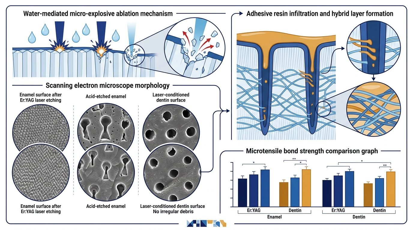
Liquid water trapped within mineralized hydroxyapatite crystals vaporizes instantaneously, expanding sixteen hundred times in volume. This volumetric expansion generates immense interstitial pressures exceeding the compressive and tensile yield limits of human enamel and dentin. Micro-explosive ejection of microcrystalline fragments occurs before significant thermal conduction reaches surrounding dental pulp. Coaxial water spray serves a dual role: it replenishes surface hydration for continuous ablation and cools irradiated substrates, maintaining pulpal chamber temperature shifts beneath two degrees Celsius.
Enamel Micro-Morphology: Prism Exposure Without Acid Demineralization
Enamel surfaces irradiated with calibrated erbium lasers display a characteristic chalky, irregular topography devoid of rotary smear debris. Microscopic examination demonstrates selective ablation of prism cores versus prism sheaths, reflecting differential water and carbonate distribution between these micro-anatomical zones. Prism boundaries remain elevated, creating macro- and micro-retentive irregularities well-suited for mechanical interlocking with polymeric resins.
Unlike thirty-seven percent phosphoric acid, which leaches calcium ions and creates deep etched microporosities fifteen to twenty micrometers deep, erbium ablation produces irregular scaly fractures without chemical dissolution. The laser-prepared enamel exhibits high acid resistance due to thermal recrystallization and carbonate depletion, a phenomenon that protects margin interfaces against secondary caries. However, resin penetration into laser-induced micro-fissures is less uniform than into acid-etched enamel unless a brief chemical conditioning step follows laser irradiation. Clinicians must recognize that physical micro-fractures behave differently from chemically dissolved prisms during adhesive polymerization.
Dentin Substrate Conditioning: Smear Layer Removal and Tubule Patency
Dentin presents an intricate organic-inorganic composite containing twenty percent collagen type one, ten percent water, and seventy percent mineralized apatite by weight. Rotary diamond or carbide burs pulverize this delicate architecture, depositing a dense smear layer containing crushed mineral particles, denatured collagen fragments, and bacteria. This smear layer occludes tubule orifices and inhibits resin monomer penetration.
Erbium laser ablation completely vaporizes the smear layer without producing demineralized collagen mats. Dentinal tubules appear completely open and patent, showing clean peritubular cuffs that protrude slightly above the surrounding intertubular dentin bed. Because peritubular dentin contains higher mineral content and less water than intertubular zones, it resists ablation, yielding a volcano-like microscopic architecture. Crucially, the exposed intertubular collagen fibrils undergo mild superficial thermal denaturant changes. Instead of soft, dangling collagen strands prone to collapse, laser-treated dentin exhibits a fused, partially denatured organic matrix that resists enzymatic hydrolysis by host-derived collagenases.
The Resin-Dentin Interface: Hybrid Layer Formation and Nano-Leakage
The altered organic state of laser-conditioned dentin dictates distinctive hybrid layer characteristics. Conventional wet bonding requires penetration of hydrophilic monomers through dense, demineralized collagen meshes. When resin monomers fail to infiltrate the full depth of acid-etched collagen, unhybridized fibrils degrade over time via endogenous matrix metalloproteinases and cathepsins, leading to adhesive failure.
In contrast, erbium-conditioned dentin produces a thinner, more densely mineralized hybrid layer measuring between one and two micrometers. Resin tags penetrate patent tubules, creating cylindrical mechanical locks. However, micro-tensile bond strength testing reveals that laser conditioning alone yields lower immediate bond values than traditional acid-etching techniques. Subsurface microcracks generated by mechanical shockwaves create micro-leakage pathways at the hybrid base. Combining low-fluence erbium conditioning with brief phosphoric acid application resolves this deficit, establishing high initial retention alongside resistance to enzymatic breakdown.
Dosimetric Parameters and Pulse Duration Optimization
Adhesion outcomes depend strictly on laser parameters, pulse duration, and water irrigation rates. Excessive pulse energy converts gentle micro-explosions into destructive mechanical shattering, producing extensive microfissures that compromise restoration margins. For enamel conditioning, clinicians should employ fluences between 12 and 16 Joules per square centimeter at 15 to 20 Hertz, using short pulse modes of fifty to one hundred microseconds. Water spray must be maintained at forty to fifty milliliters per minute.
Dentin conditioning requires conservative fluences between 6 and 10 Joules per square centimeter with pulse durations around one hundred microseconds. Long pulses allow thermal accumulation, causing carbonization, collagen burning, and complete loss of bond capacity. Calibrating tip-to-tissue distance between one and two millimeters under perpendicular orientation ensures uniform energy deposition across cavity preparations.
Comparative Longevity and Clinical Bond Durability
Long-term in vitro aging studies, including thermal cycling and mechanical fatigue simulations, reveal distinct performance characteristics between laser-prepared and rotary-prepared restorations. While conventional acid-etched dentin interfaces lose up to forty percent of micro-tensile bond strength after twelve months of water storage, erbium-prepared interfaces demonstrate stable bond metrics. Resistance to hydrolytic degradation stems from the absence of deep denuded collagen zones.
Clinical trials over five years confirm that class five cervical restorations prepared with erbium lasers exhibit retention rates exceeding ninety-two percent, equivalent to conventional adhesive preparations. Margins prepared with erbium lasers display significantly lower rates of marginal discoloration and recurrent decay, attributed to thermal mineral alteration and lack of marginal gap expansion. Combining laser cavity preparation with modified adhesive protocols capitalizes on biological preservation while securing durable seal integrity.
Practical Synthesis for Restorative Dentistry
Erbium lasers represent an advanced hard-tissue conditioning tool, not a universal replacement for adhesive chemistry. Clinicians who understand the biophysical differences between thermal micro-ablation and acid dissolution achieve superior restorative longevity. By tailoring fluence, pulse duration, and supplementary conditioning, the dental practitioner eliminates smear debris, preserves natural dentin mineral, and establishes stable resin adhesion resistant to hydrolytic degradation.
References
- De Moor, R. J., et al. (2010). Erbium lasers in endodontics and restorative dentistry. Lasers in Medical Science, 25(6), 889-897.
- Dunn, W. J., et al. (2005). Microtensile bond strength of composite resin to Er:YAG-laser-prepared enamel and dentin. The Journal of the American Dental Association, 136(5), 622-626.
- Cardoso, M. V., et al. (2008). Influence of Er,Cr:YSGG laser on the micro-tensile bond strength of adhesive systems to dentin. Journal of Dentistry, 36(6), 444-452.
- Walsh, L. J. (2003). The current status of laser applications in dentistry. Australian Dental Journal, 48(3), 146-155.
- Ramos, A. C., et al. (2014). Adhesion to Er:YAG laser-prepared dentin: a review of the literature. Photomedicine and Laser Surgery, 32(8), 430-438.
Leave a Reply