

The erbium-doped yttrium aluminum garnet laser operating at 2,940 nanometers enables selective ablation of infected dentin while preserving underlying affected collagen matrix and maintaining pulpal thermal safety through synchronized water-spray cooling. Cavity preparation using conventional high-speed diamond burs and low-speed carbide burs has defined restorative dentistry for over a century. However, rotary mechanical cutting relies on frictional shearing, which generates substantial microvibrations, auditory discomfort, friction heat, and microcracks within surrounding enamel and dentinal tubules. Furthermore, rotary burs invariably produce an amorphous smear layer that plugs dentinal tubules and weakens adhesive resin monomer infiltration. The 2,940 nm Er:YAG laser circumvents these mechanical drawbacks by utilizing thermomechanical micro-explosions that ablate demineralized tissue selectively while opening dentinal tubules without producing an inorganic smear layer.
Optical Physics and Differential Water Absorption Mechanics
The primary mechanism underlying Er:YAG laser hard tissue ablation resides in coincidence with the major absorption peak of liquid water at 2.94 micrometers. At this wavelength, the optical absorption coefficient in water exceeds 10,000 cm-1, surpassing carbon dioxide and diode wavelengths by orders of magnitude. Hydroxyapatite also exhibits a secondary phosphate-hydroxyl absorption band near 2.8 to 3.0 micrometers.
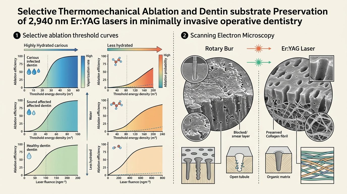
When short laser pulses (100 to 300 microseconds) irradiate hydrated dentin, photonic energy is absorbed instantaneously by interstitial water trapped within the mineralized collagen matrix. The absorbed radiation causes explosive volumetric expansion of liquid water into superheated steam within nanoseconds. This violent phase transition generates localized pressure gradients exceeding the tensile strength of the surrounding inorganic mineral scaffold, causing micro-explosive ejection of microscopic mineral fragments. Because carious, demineralized dentin possesses higher water content (often exceeding 25% to 35% by volume) and lower mineral density than sound dentin (approx. 10% water), the Er:YAG laser ablates infected dentin at a substantially lower energy threshold than healthy dentin, conferring natural biological selectivity.
Selective Dentin Ablation: Preserving the Affected Dentin Zone
Modern operative principles mandate distinguishing between the outer layer of carious dentin (caries-infected dentin) and the inner layer (caries-affected dentin). Caries-infected dentin is irreversibly denatured, densely infiltrated by polymicrobial biofilms, devoid of remineralizable crystal seeds, and mechanically unviable; it must be completely excised. In contrast, caries-affected dentin retains intact intermolecular cross-linked collagen scaffolding and mineralizable apatite crystals, serving as an ideal biological base for remineralization and pulpal protection.
With mechanical round burs, tactile distinction between infected and affected dentin remains subjective, frequently leading to over-excavation and accidental pulpal exposure in deep lesions. By adjusting Er:YAG laser fluence to between 8 and 12 J/cm2 (pulse energies of 120 to 180 mJ at 15 to 25 Hz), ablation self-terminates when the beam transitions from moist, soft infected tissue to firmer, less hydrated affected dentin. The practitioner perceives a distinct auditory shift from a dull, muffled pop to a sharp, crisp clicking sound, accompanied by resistance to further cavitation. This acoustic feedback provides an objective physical boundary that prevents inadvertent pulpal fenestration.
Pulpal Thermal Safety and Hemodynamic Response
Protecting the vital dental pulp from irreversible thermal injury represents the foremost safety parameter during laser cavity preparation. Seminal investigations by Zach and Cohen demonstrated that an intrapulpal temperature elevation of 5.5 degrees Celsius produces irreversible pulpal necrosis in 15% of teeth, while an elevation of 11.1 degrees Celsius causes total pulpal necrosis in 100% of cases.
The Er:YAG laser preserves intrapulpal temperature stability through continuous coaxial water-air spray. Delivering sterile water at rates between 3.5 and 5.0 mL/min across the focal target maintains average pulpal temperature fluctuations within 1.5 to 2.5 degrees Celsius below baseline levels. The water film acts simultaneously as an essential photon absorber for the ablation process and a constant thermal heat sink that absorbs excess convective heat. Furthermore, because thermal diffusion is restricted by the microsecond pulse duration, adjacent pulpal tissue shows no microvascular thrombosis, disorganization of the odontoblastic layer, or neutrophil infiltration in histological animal evaluations.
Ultrastructural Morphology: Smear Layer Elimination and Hybrid Layer Formation
Scanning electron microscopy (SEM) reveals striking ultrastructural differences between surfaces prepared by rotary burs and those ablated by Er:YAG laser. Rotary burs burnish necrotic debris into enamel and dentin, creating a 2 to 5 micrometer thick smear layer with dense smear plugs obliterating tubule orifices. Removing this smear layer requires separate acid etching with 37% phosphoric acid, which can over-demineralize deep dentin and expose collagen to matrix metalloproteinases (MMPs).
In contrast, Er:YAG ablated dentin exhibits a clean, irregular, micro-retentive surface devoid of any smear layer. Dentinal tubules remain patent and funnel-shaped, free of debris. Intertubular dentin is selectively ablated more rapidly than peritubular dentin due to differential water and mineral distribution, creating a micro-scalloped topography that enhances micromechanical interlocking of adhesive resins. However, laser ablation produces a thin (0.5 to 1.5 micrometer) sub-surface microfissured zone containing chemically altered, partially fused collagen. Clinicians must adjust their adhesive protocol: applying universal self-etch adhesives or mild phosphoric acid conditioning for five to ten seconds cleanses this altered surface layer, allowing resin monomers to infiltrate open tubules and form robust hybrid layers with minimal microleakage.
Patient Experience and Reduced Local Anesthetic Requirement
A pronounced clinical advantage of Er:YAG laser cavity preparation is the elimination of mechanical vibration and bone-conducted bone resonance. Mechanical bur rotation at 300,000 to 400,000 RPM generates intense microvibrations that propagate through cranial bones directly to the inner ear, triggering deep somatic anxiety.
Because laser ablation operates without mechanical contact, pressure-sensitive dentinal mechanoreceptors and intradental hydrodynamic fluid shifts remain largely undisturbed. The acoustic pop of laser pulses induces slight pulpal desensitization via transient hyperpolarization of unmyelinated nerve terminals. Over 80% of small to medium class I, class III, and class V cavity preparations can be completed without local anesthetic injections, eliminating needle phobia, prolonged lip numbness, and postoperative masticatory soft tissue trauma.
Clinical Guidelines and Best Practices
To maximize clinical efficiency while preventing surface desiccation, practitioners should adhere to specific operational rules. The laser tip must be held at a 90-degree angle to the target surface at a working distance of 0.5 to 1.0 millimeters in non-contact mode, moving continuously in cross-hatch strokes. Stagnant positioning over a single spot produces localized deep grooving. Water flow must never be interrupted during active emission. By mastering these biophysical fundamentals, restorative dentists elevate operative precision, preserve natural tooth structure, and optimize long-term restoration longevity.
References
- Aoki, A., et al. (2004). Current status of Er:YAG laser in restorative dentistry: a review. Journal of Clinical Laser Medicine & Surgery, 22(3), 209-216.
- Coluzzi, D. J. (2004). Fundamentals of dental lasers: science and instruments. Dental Clinics of North America, 48(4), 751-770.
- De Moor, R. J., et al. (2010). The effect of Er:YAG laser preparation on the microleakage of composite restorations. Lasers in Medical Science, 25(6), 843-850.
- Hibst, R., & Keller, U. (1989). Experimental studies of the application of the Er:YAG laser on dental hard substances: I. Measurement of the ablation rate. Lasers in Surgery and Medicine, 9(4), 338-344.
- Zach, L., & Cohen, G. (1965). Pulp response to externally applied heat. Oral Surgery, Oral Medicine, Oral Pathology, 19(4), 515-530.
Leave a Reply