
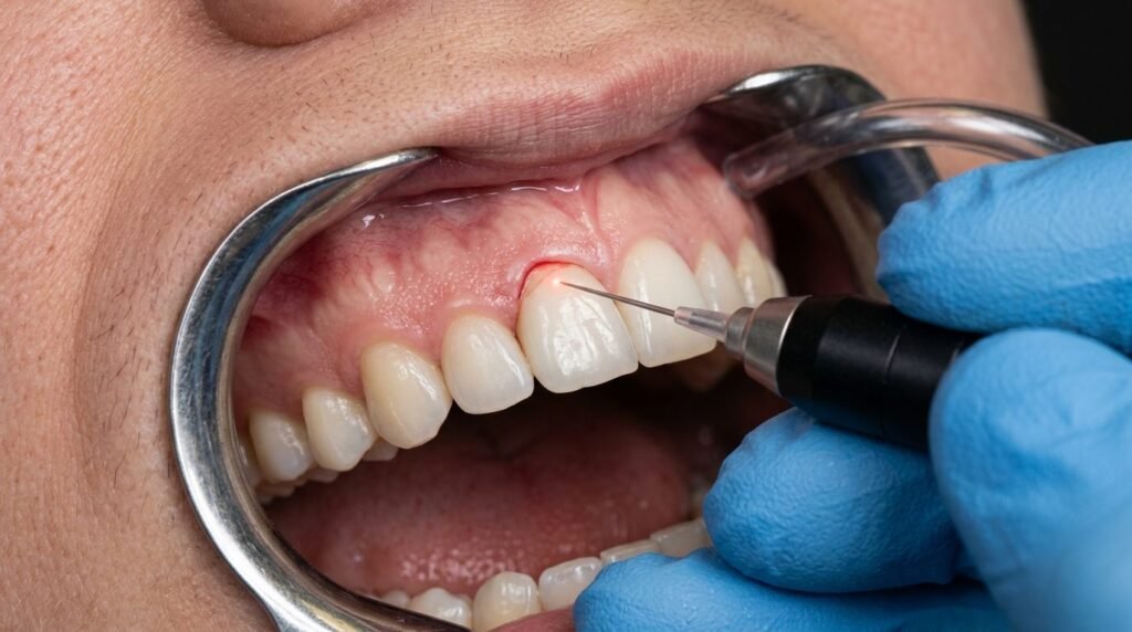
Surgical excision of oral soft tissues has historically relied on steel scalpels or high-frequency electrosurgery units. While conventional scalpels offer clean incisions with minimal lateral tissue damage, profuse capillary hemorrhage obscures the surgical field and necessitates sutures. Electrosurgery achieves hemostasis through electrical resistance heating but risks uncontained thermal conduction into underlying alveolar bone and periosteum. In contrast, high-power near-infrared diode lasers operating at 810 nm, 940 nm, and 980 nm provide precise photothermal incision combined with instantaneous microvascular coagulation. Light energy guided through fine quartz fibers vaporizes intracellular water while sealing capillaries smaller than 0.5 millimeters in diameter. However, improper tip initiation, excessive power densities, or prolonged dwell times generate deep lateral carbonization zones that delay secondary intention epithelialization. Understanding optical physics, thermal relaxation times, and biological damage boundaries remains essential for optimizing surgical outcomes during gingivectomy and frenectomy procedures.
Optical Absorption Characteristics of 810, 940, and 980 nm Wavelengths
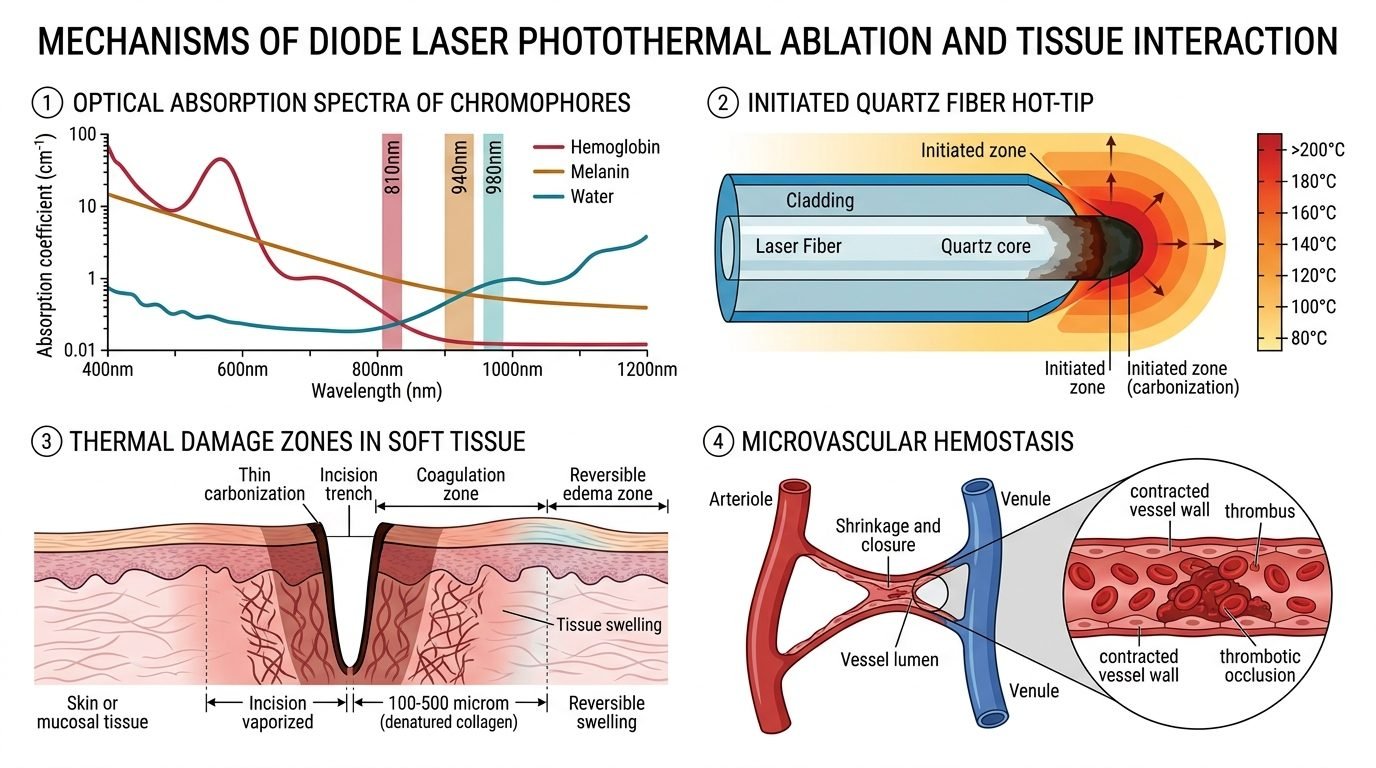
Diode lasers belong to the class of solid-state semiconductor lasers that emit coherent radiation in the near-infrared spectrum. Biological soft tissues lack significant water absorption within this optical range. Instead, photothermal ablation relies on optical absorption by endogenous chromophores, primarily oxyhemoglobin, deoxyhemoglobin, and melanin. The 810 nm wavelength aligns closely with the peak absorption spectrum of deoxygenated hemoglobin, making it well-suited for hyperemic or highly vascularized granulomatous lesions.
The 940 nm wavelength exhibits balanced absorption between hemoglobin and secondary water peaks, providing efficient soft-tissue cutting efficiency at lower power inputs. The 980 nm wavelength demonstrates slightly higher water absorption than 810 nm, improving superficial vaporization while maintaining moderate hemostatic capability. Because near-infrared light scatters deeply within unpigmented collagenous connective tissue, the bare uninitiated fiber behaves as an optical radiator rather than a focused cutting knife. Achieving precise scalpel-like incisions requires converting this optical transmission into localized contact photothermal energy at the fiber tip.
Fiber Initiation, Hot-Tip Effects, and Photothermal Incision
To cut oral soft tissue efficiently without uncontained forward radiation, clinicians must initiate the distal fiber tip. Fiber initiation involves coating the cleaved quartz tip with an organic articulating carbon film, cork, or blood pigment. When the laser fires, the dark pigment absorbs near-infrared photons instantaneously, elevating tip temperature to between 500 and 800 degrees Celsius. The fiber tip transforms into an intensely heated optothermal cutting instrument, colloquially termed a hot-tip.
During tissue contact, incandescent heat transfers directly to target cells, causing flash boiling of intracellular water and immediate structural carbonization. As steam expands rapidly, cell membranes rupture, separating tissue along the path of fiber movement. Concurrently, unabsorbed residual photons penetrate forward into adjacent capillary loops, denaturing endothelial proteins and activating the coagulation cascade. Microvascular thrombosis seals arterioles and venules, providing an entirely bloodless operative field. This bloodless environment improves visualization during delicate aesthetic gingivoplasties and eliminates the need for postsurgical suturing in labial or lingual frenectomies.
Lateral Thermal Damage Boundaries and Coagulation Zones
Histological examinations comparing scalpel, electrosurgery, and diode lasers demonstrate distinct wound margins. Scalpel incisions produce zero lateral thermal necrosis, allowing primary intention healing with minimal myofibroblastic contraction. High-power diode lasers generate a layered thermal injury profile: an inner vaporized trench, surrounded by a thin layer of carbonized tissue, followed by a zone of irreversible thermal coagulation spanning 100 to 500 microns. Beyond this coagulation boundary lies a reversible sub-lethal zone characterized by temporary cellular edema and vascular hyperaemia.
The width of the lateral coagulation zone depends strictly on surgical technique and laser emission parameters. Continuous wave emission causes heat accumulation within surrounding connective tissue because energy delivery exceeds the biological thermal relaxation time. Pulsed emission modes, such as gated or microsecond pulse trains, provide essential cooling intervals between bursts, limiting thermal diffusion to less than 200 microns. Clinicians must maintain a continuous light brushing movement at approximately two to three millimeters per second, cooling the operative site with gentle air or saline irrigation.
Gingivectomy and Aesthetic Margin Repositioning
In aesthetic gingivectomies and crown lengthening procedures, diode lasers offer unparalleled margin sculpting precision. Orthodontic patients exhibiting drug-induced or plaque-induced gingival hyperplasia benefit from bloodless excision that permits immediate bracket bonding or impression taking. The clinician uses an initiated 300 to 400 micron fiber tip angled at 45 degrees to the long axis of the tooth, beveling the gingival margin toward the enamel surface.
Maintaining a minimum distance of two to three millimeters from the alveolar crest protects the biological width. Applying high-power diode energy directly against the periosteum or marginal alveolar crest causes localized osteonecrosis, bony sequestration, and persistent gingival recession. Using low average powers between 0.8 and 1.2 Watts in pulsed mode prevents periosteal heat transmission. Because the diode laser seals peripheral lymphatic channels alongside capillaries, postoperative swelling is markedly diminished compared to conventional periodontal flap surgery.
Lingual and Labial Frenectomy Protocols
Ankyloglossia and aberrant maxillary labial frenula represent ideal indications for diode laser excision. In pediatric and infant tongue-tie release, eliminating general anesthesia and suturing transforms a stressful hospital intervention into an outpatient procedure. Under minimal topical or localized infiltration anesthesia, the clinician elevates the tongue with a grooved director, exposing the fibrotic fascia of the genioglossus muscle.
The initiated fiber tip sweeps gently in horizontal strokes, releasing fibrous bands layer by layer until unrestricted lingual mobility is verified. The surgical diamond-shaped defect heals by secondary intention under a protective fibrinous coagulum that forms immediately post-excision. This autologous fibrin seal acts as a biological dressing, shielding unmyelinated nerve endings from salivary enzymes and food debris. Consequently, patients report minimal postoperative discomfort and rarely require analgesic medication beyond simple acetaminophen.
Postoperative Healing Kinetics and Complication Management
Secondary intention healing following diode laser surgery exhibits a slightly delayed initial epithelialization phase compared to scalpel incisions. The thin layer of denatured collagen and coagulated protein must be cleared by host macrophages before basal keratinocytes migrate across the wound bed. Complete re-epithelialization typically requires 10 to 14 days, culminating in a smooth, scarless mucosal architecture due to reduced myofibroblastic proliferation.
Complications arise almost exclusively from operator technical errors, primarily excessive power settings and static fiber positioning. Thermal necrosis of underlying periosteum or cementum produces localized sloughing, severe pain, and delayed healing. Preventing these adverse events requires disciplined adherence to lowest-effective-power principles, adequate tip cleaning with sterile damp gauze, and constant fiber motion. When managed with biological understanding, high-power diode lasers deliver exceptional surgical precision and unmatched patient comfort.
References
- Romanos, G. E. (2004). Diode laser soft-tissue surgery. Compendium of Continuing Education in Dentistry, 25(12), 922-928.
- Goharkhay, K., et al. (1999). Soft tissue surgery in the oral cavity with a 940 nm diode laser. Lasers in Surgery and Medicine, 25(5), 416-422.
- Cobb, C. M. (2006). Lasers in periodontics: a review of the literature. Journal of Periodontology, 77(4), 545-564.
- Fornaini, C., et al. (2012). 810 nm, 980 nm, and 1064 nm laser irradiation of oral soft tissues: An in vitro comparative study. Journal of Clinical and Diagnostic Research, 6(8), 1439-1443.
- Amaechi, B. T. (2019). Dental Erosion and Its Clinical Management. Springer Nature.
Leave a Reply