

A severe gag reflex in the dental operatory is not a behavioral tantrum; it is an unconditioned protective brainstem motor program designed by evolution to prevent foreign body asphyxiation.
Patients plagued by an exaggerated gag reflex dread dental visits as intensely as phobics fear needles. When dental instruments touch the posterior tongue or soft palate, their abdominal wall contracts violently. Retching, coughing, lacrimation, and tachycardia erupt in rapid, uncontrollable succession. Clinicians often instruct the patient to relax or dismiss it as voluntary exaggeration. This clinical attitude reflects profound neurological ignorance. The gag reflex is governed by primitive autonomic nuclei in the medulla oblongata that fire hundreds of milliseconds before conscious cognitive deliberation can intervene.
The physical consequence is diagnostic and restorative paralysis. Radiographs are abandoned, margins remain unsealed, and impressions trigger choking episodes. Over time, patients avoid clinics out of humiliation and physiological exhaustion. Managing this reflex requires abandoning frustration and applying targeted neurosensory gating, pharmacological inhibition, and proprioceptive distraction.
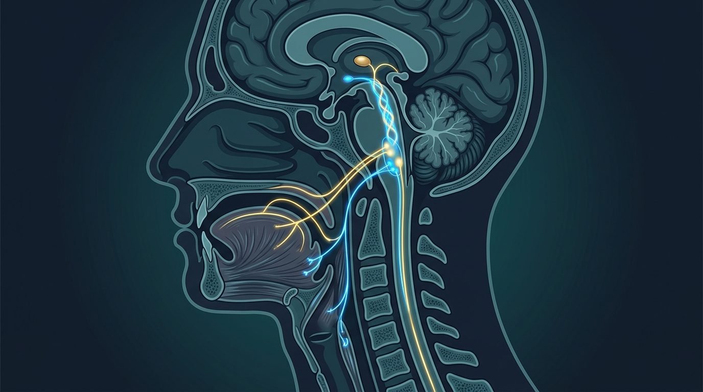
The Neuroanatomical Circuitry of the Pharyngeal Reflex
The gag reflex constitutes an involuntary motor arc orchestrated through multiple cranial nerves. Mechanical tactile stimulation of five primary trigger zones initiates the afferent limb of the circuit: the posterior third of the tongue, the palatoglossal and palatopharyngeal arches, the soft palate, the uvula, and the posterior pharyngeal wall.
Sensory afferents travel primarily via the glossopharyngeal nerve (cranial nerve IX), with minor contributions from the trigeminal (cranial nerve V) and vagus (cranial nerve X) nerves. These primary sensory axons project directly into the solitary tract nucleus and the spinal trigeminal nucleus within the brainstem.
Interneurons integrate signals in the medullary reticular formation. This complex connects immediately to the bilateral nucleus ambiguus and the hypoglossal nucleus (cranial nerve XII).
The efferent response fires through the vagus nerve and glossopharyngeal branches. Motor impulses cause simultaneous elevation of the soft palate to seal the nasopharynx, superior pharyngeal constrictor contraction to close the pharyngeal lumen, and diaphragm-abdominal muscle spasm to expel foreign matter upward.
Cortical and limbic inputs modulate this medullary center. In patients with psychomotor gagging, visual perception of a dental tray or the olfactory stimulus of alginate powder activates the basolateral amygdala, firing descending facilitating tracts that lower the firing threshold of the nucleus ambiguus to zero. The reflex fires before physical contact occurs.
Anatomy of the Medullary Gag Reflex Arc:
- Afferent Pathway: Glossopharyngeal nerve (CN IX) carrying mechanical tactile signals from posterior oral mucosa.
- Central Integration Center: Solitary tract nucleus and medullary reticular formation in the medulla oblongata.
- Efferent Pathway: Vagus nerve (CN X) and hypoglossal nerve (CN XII) driving pharyngeal constriction and tongue retraction.
- Sensory Modulators: Descending corticobulbar and amygdaloid tracts mediating anticipatory psychomotor nausea.
The Neurobiology of Sensory Gating and Proprioceptive Distraction
The central nervous system cannot process infinite high-intensity inputs simultaneously. Gate control theory, originally formulated for spinal pain gating, applies directly to brainstem cranial nerve integration.
Flooding competing somatosensory pathways dampens glossopharyngeal transmission. When a patient actively contracts large voluntary muscle groups distant from the head, motor cortex activity consumes significant central processing bandwidth.
Instructing the patient to raise one leg straight off the dental chair and hold it rigidly suspended forty-five degrees above horizontal activates the femoral quadriceps and core abdominal stabilizers. Motor neurons in the precentral gyrus discharge sustained action potentials. The reticular formation prioritizes proprioceptive and motor feedback from the lower extremity, dampening background pharyngeal reflex sensitivity.
Holding table salt on the anterior tongue tip provides sensory distraction. Placing a few granules of sodium chloride on the tip of the tongue stimulates taste receptor cells innervated by the chorda tympani (cranial nerve VII). Gustatory afferents ascend to the rostral solitary nucleus, where they trigger local inhibitory interneurons that attenuate glossopharyngeal reflex sensitivity across the soft palate for several minutes.
“Telling a gagging patient to just relax is as scientifically useless as telling an asthmatic to breathe deeper; the brainstem reflex fires before conscious thought can intervene.”
Forced nasal respiration alters airway mechanics. Mouth breathing drops the soft palate onto the tongue base, maximizing mucosal friction against dental instruments. Continuous rhythmic inhalation and exhalation through the nose elevates the velum and stabilizes the uvula, widening the functional oropharyngeal corridor.
Acupressure at Pericardium 6 (Neiguan) and Autonomic Control
Acupoint stimulation provides measurable anti-emetic neuro-modulation. The Pericardium 6 (P6 or Neiguan) acupoint, located on the anterior forearm two proportional finger-breadths proximal to the wrist crease between the tendons of palmaris longus and flexor carpi radialis, is supported by extensive biomedical literature.
Controlled mechanical pressure on P6 stimulates sensory fibers of the median nerve. Afferent median nerve impulses project to the nucleus tractus solitarius and hypothalamus, promoting central beta-endorphin release and suppressing emetic motor outputs from the area postrema.
Applying elastic acupressure wristbands featuring rigid plastic hemispherical studs bilaterally fifteen minutes prior to procedure commencement reduces gagging severity scores by over fifty percent in randomized controlled clinical trials.
Ear acupressure at the vagal auricle provides supplementary inhibition. Stimulating the cymba conchae, the cutaneous zone innervated by the auricular branch of the vagus nerve (Arnold’s nerve), activates central vagal parasympathetic nuclei, slowing heart rates and calming anticipatory retching.
Clinical Protocol for Restorative and Impression Success
Clinical pacing requires systematic restructuring. Impression taking represents the ultimate challenge. Clinicians must abandon oversized metal trays and runny, slow-setting alginates.
Digital intraoral scanning eliminates bulk material completely. Scanning allows immediate pausing. The wand tip can rest against buccal mucosa while the patient swallows, eliminating the claustrophobic dread of tray entrapment.
When physical impressions are unavoidable, fast-setting materials should be selected. The tray must be custom-scalloped to prevent excess material from pooling onto the soft palate. The patient sits completely upright with their chin tilted downward, preventing saliva from pooling into the posterior pharynx.
Topical local anesthetics applied to the soft palate provide chemical receptor dampening. Viscous two percent lidocaine painted over palatoglossal arches blocks sensory mechanoreceptors for fifteen minutes, allowing diagnostic procedures to conclude smoothly.
Understanding the neurobiology of the gag reflex converts operatory chaos into clinical composure. Respecting brainstem physiology preserves patient dignity and guarantees high-level restorative outcomes.
Systematic sensory gating, P6 acupressure, and upright patient positioning resolve severe pharyngeal retching without recourse to hospital general anesthesia.
Scientific References and Supporting Literature
- Winocur, E., Emodi-Perlman, A., Friedman-Rubin, P., et al. (2011). The association between severe gag reflex and dental anxiety. Journal of Public Health Dentistry, 71(4), 312-317.
- Bassi, G. S., Humphris, G. M., & Longman, L. P. (2004). The etiology and management of gagging: a review of the literature. The Journal of Prosthetic Dentistry, 91(5), 459-467.
- Dickinson, C. M., & Fiske, J. (2005). A review of gagging problems in dentistry: 1. Aetiology and classification. British Dental Journal, 199(1), 26-29.
- Sari, E., & Sari, T. (2010). The role of acupuncture in the management of severe gag reflex: a case report. Acupuncture in Medicine, 28(4), 211-213.
- Lu, D. P., & Lu, G. P. (2012). Clinical management of severe gag reflex in dental patients: a review and report of cases. Compendium of Continuing Education in Dentistry, 33(10), 738-744.
Leave a Reply