
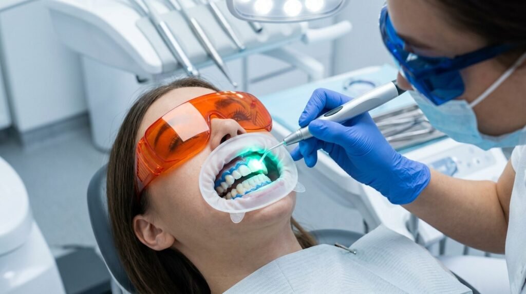
Dental bleaching lights that heat tooth enamel are obsolete radiative heaters; true laser photo-activation accelerates peroxide breakdown through narrow-band photochemical excitation while keeping intrapulpal temperatures well below biological necrosis thresholds.
For over two decades, in-office cosmetic dentistry relied on broad-spectrum plasma arc lamps, ultraviolet emitters, and generic light-emitting diode arrays. These non-coherent sources emit vast quantities of unfocused polychromatic radiation spanning broad electromagnetic wavebands. Water molecules within enamel and dentin absorb this incidental energy, transforming light into brute heat. The pulp chamber experiences severe thermal stress. Patients endure agonizing intraoperative pulpal zings, followed by days of debilitating postoperative thermal hypersensitivity. Many clinicians concluded that light activation was an aggressive marketing gimmick that inflicted unnecessary pulpal trauma without improving aesthetic longevity.
That critique accurately describes indiscriminate radiative heating, but it mischaracterizes true laser physics. Coherent monochromatic laser radiation operates through targeted photon-matter interactions. When narrow-band laser wavelengths match specific photo-initiators or reactive oxidation absorption bands within bleaching gels, photochemical peroxide cleavage accelerates without elevating internal pulpal temperature. The difference between crude thermal acceleration and laser-assisted bleaching is the difference between scorched enamel and refined optical photochemistry.
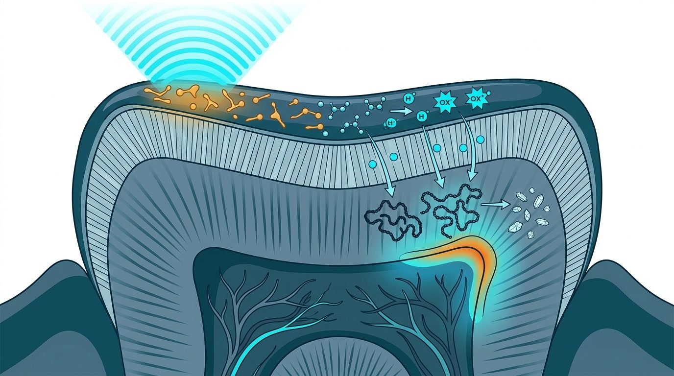
Photochemical Decomposition Versus Bulk Thermal Degradation
Hydrogen peroxide bleaches discolored substrates through the generation of reactive oxygen species. Hydroxyl free radicals, perhydroxyl anions, and superoxide radicals oxidize complex organic chromophores locked within the mineralized inter-prismatic enamel matrix.
Long, conjugated carbon ring double-bonds absorb visible light and cast dark yellow-brown hues. Reactive oxygen species cleave these conjugated bonds, converting dark pigments into simpler, non-pigmented hydrophilic chains that reflect light cleanly. The process restores youthful tooth translucency without dissolving underlying hydroxyapatite prisms.
Generic bleaching lamps accelerate this oxidation by raising overall gel temperature. According to standard thermodynamic principles, every ten-degree Celsius rise in temperature doubles the rate of chemical reactions. That temperature increase transfers straight through enamel and dentin into the pulp chamber, threatening microvascular collapse.
Specific laser wavelengths circumvent bulk heating. Diode lasers emitting at 810 nanometers or 980 nanometers, and potassium-titanyl-phosphate (KTP) lasers emitting at 532 nanometers, interact with tailored chromophores suspended inside specialized bleaching formulations.
Photon energy is absorbed almost entirely within the chemical gel layer itself. The peroxide molecules undergo rapid dissociation into perhydroxyl radicals via resonant vibrational activation while the underlying mineralized tooth structure remains physiologically cool.
Optical and Thermal Metrics in Power Bleaching:
- Critical Pulpal Threshold: Maximum safe temperature rise is delta T of 5.5°C before irreversible pulpitis occurs.
- Conventional LED/Plasma Lamps: Intrapulpal temperature surges frequently range from 6.0°C to 12.0°C.
- Calibrated KTP/Diode Lasers: Mean intrapulpal temperature rise remains strictly controlled between 1.1°C and 2.4°C.
- Enamel Microhardness: Surface mineral density preserved without acid-induced prismatic dissolution or pitting.
The Critical 5.5-Degree Pulpal Ceiling and Calorimetric Tracking
The dental pulp is an exquisitely vascularized organ confined within rigid, non-compliant dentinal walls. It possesses zero collateral circulation and minimal tolerance for elevated thermal environments.
Seminal thermal threshold studies established that an intrapulpal temperature increase of 5.5 degrees Celsius causes irreversible histological pulp necrosis in fifteen percent of teeth. A rise of eleven degrees Celsius produces complete pulpal necrosis in one hundred percent of cases.
Standard halogen and plasma curing lights regularly produce intrapulpal temperature surges exceeding seven to ten degrees Celsius when directed continuously at labial surfaces for twenty minutes. The pulp suffers acute microvascular stasis, thrombosis, and subsequent sterile coagulative necrosis.
Calibrated diode and KTP lasers maintain tight thermal profiles. Thermocouple investigations confirm that pulsed diode laser applications deliver sufficient photonic flux to split peroxide bonds while maintaining mean intrapulpal temperature increases below two degrees Celsius. The pulp remains in a safe biological zone. Postoperative inflammatory pulpal hyperemia is avoided, eliminating the severe spontaneous spasms that haunt patients after conventional light bleaching.
“Broad-spectrum bleaching lamps bake the dental pulp to speed up peroxide; monochromatic lasers activate the chemistry inside the gel while the tooth remains cool.”
Exposure times are dramatically shortened. Because photochemical reaction kinetics proceed rapidly under coherent irradiation, total chairside peroxide contact time drops from sixty minutes to less than fifteen minutes. Reduced exposure duration translates into less chemical diffusion through dentinal tubules toward the pulp horn.
Preservation of Enamel Ultrastructure and Microhardness
Extended exposure to acidic hydrogen peroxide formulations degrades enamel surface integrity. Commercial bleaching gels formulated at low pH levels to ensure shelf stability can etch prism cores and lixiviate calcium and phosphate ions from hydroxyapatite lattices.
Scanning electron microscopy of enamel exposed to prolonged conventional bleaching reveals surface pitting, prismatic irregularities, and increased roughness. This altered surface morphology acts as a microscopic trap that attracts dietary tannins and chromogenic bacteria, causing rapid shade rebound within months.
Laser-activated protocols protect mineral density. Because total chemical contact time is slashed by seventy percent, superficial calcium loss is minimized.
Vickers microhardness testing shows that enamel treated with laser-activated bleaching retains baseline surface microhardness comparable to untreated control surfaces. Prismatic morphology remains smooth, preventing post-treatment plaque accumulation and preserving long-term aesthetic stability.
Clinical Protocol for Predictable Laser Bleaching
Clinical predictability depends on meticulous gingival isolation and precise energy titration. The clinician must inspect enamel surfaces for microcracks, exposed dentin margins, or abfraction lesions prior to gel placement.
Light-cured resin liquid dams must be placed circumferentially around cervical margins to prevent chemical soft tissue burning. The laser-specific bleaching gel is mixed fresh to ensure peak reactive radical availability.
The laser handpiece is swept across the dental arch in non-contact scanning mode. Energy densities must not exceed recommended manufacturer thresholds. Continuous stationary irradiation of a single tooth must never occur.
Following two brief activation cycles of thirty to forty-five seconds per quadrant, the gel is aspirated completely without water spraying. Remineralizing pastes containing amorphous calcium phosphate or nano-hydroxyapatite are applied immediately to seal surface microporosities. This post-bleaching mineral bath guarantees immediate desensitization and locks in stable enamel brightness.
True laser bleaching marries aesthetic desires with pulp-protective biophysics. It demonstrates that cosmetic outcomes need not compromise pulpal vitality.
Monochromatic laser bleaching achieves superior shade improvement, maintains normal intrapulpal temperatures, and preserves prismatic enamel structure.
Scientific References and Supporting Literature
- Zach, L., & Cohen, G. (1965). Pulp response to externally applied heat. Oral Surgery, Oral Medicine, Oral Pathology, 19(4), 515-530.
- Buchalla, W., & Attin, T. (2007). External bleaching therapy with activation by heat, light or laser: a systematic review. Dental Materials, 23(5), 586-596.
- Sulieman, M. (2005). An overview of bleaching techniques: 2. Nightguard vital bleaching and non-vital bleaching. Dental Update, 32(1), 39-46.
- Dostalova, T., Jelinkova, H., Housova, D., et al. (2004). Diode laser-activated tooth bleaching. Brazilian Dental Journal, 15(Spec No), 3-8.
- Kwon, S. R., & Wertz, P. W. (2015). Review of the mechanism of tooth whitening. Journal of Esthetic and Restorative Dentistry, 27(5), 240-257.
Leave a Reply