

Traditional diagnostic reliance on sharp metal explorers and bitewing radiographs fails to detect incipient enamel demineralization before irreversible cavitation occurs. For decades, clinicians utilized forceful explorer probing into anatomical pits and fissures as the standard method for tactile caries assessment. Contemporary micro-computed tomography and histology demonstrate that mechanical pressure from an explorer fractures weakened enamel prisms over remineralizable sub-surface lesions, inadvertently creating irreversible iatrogenic cavities. Furthermore, conventional dental radiography exhibits notoriously poor sensitivity for early occlusal and smooth-surface lesions, revealing mineral loss only after demineralization penetrates well into middle coronal dentin. Quantitative laser fluorescence systems operating at 655 nm and cross-sectional optical coherence tomography provide non-destructive photonic modalities capable of quantifying subclinical mineral loss objectively, enabling minimally invasive preventive remineralization before restorative intervention becomes mandatory.
Photophysical Mechanisms of Laser Fluorescence (DIAGNOdent)
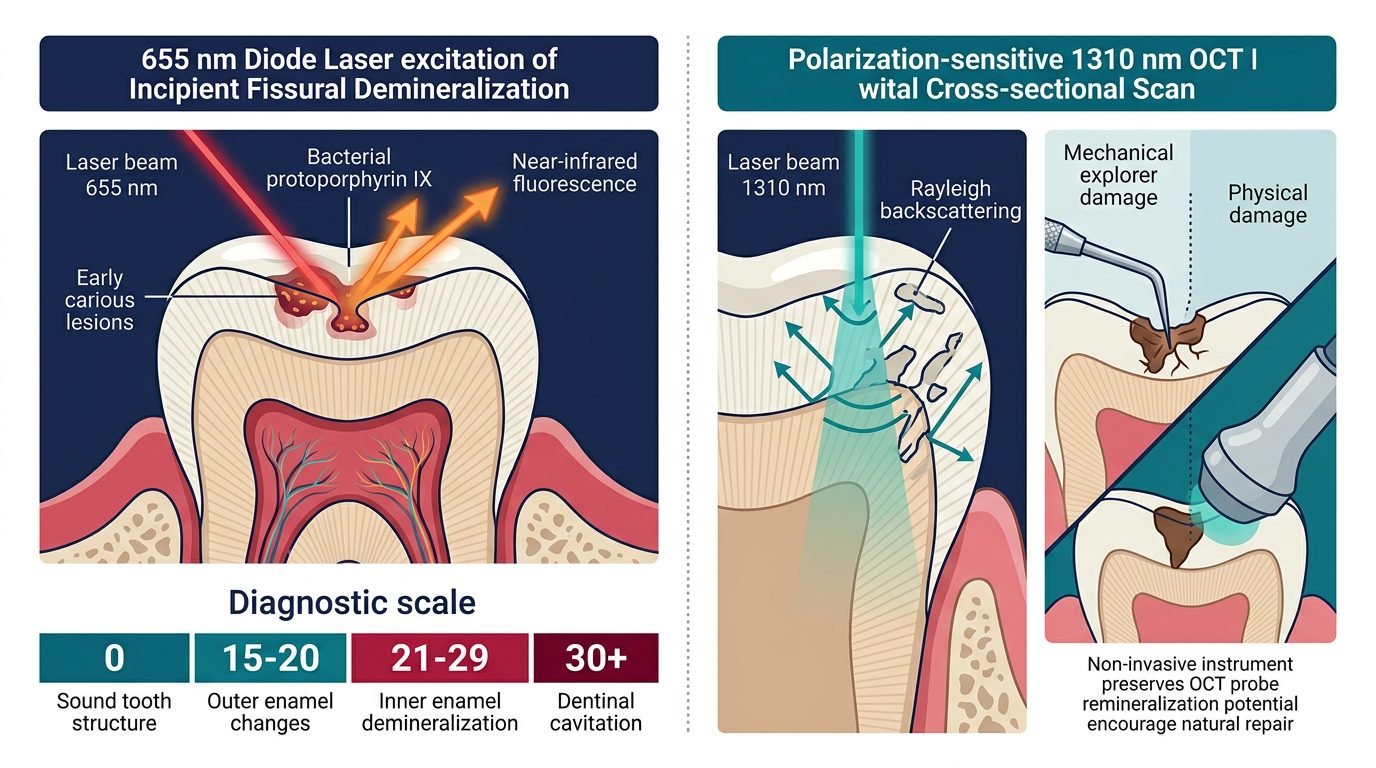
Quantitative laser fluorescence utilizes a compact semiconductor diode emitting monochromatic red light at a calibrated wavelength of 655 nanometers. This photonic emission is delivered to tooth surfaces through specialized flat or conical optical fiber tips. Unlike sound enamel, which exhibits negligible intrinsic fluorescence when exposed to red wavelengths, carious dental tissues fluoresce intensely in the near-infrared spectrum between 700 and 800 nanometers. This fluorescence does not originate from altered hydroxyapatite crystals themselves, but rather from endogenous porphyrins and protoporphyrin IX synthesized by anaerobic bacterial metabolic activity within active biofilm communities.
When the 655 nm photon beam excites these microbial fluorophores, the device’s optical detector collects backscattered light, filters out the primary 655 nm excitation wavelength, and quantifies long-wavelength infrared emissions on an arbitrary numerical scale from 0 to 99. Numerical values correlate precisely with bacterial colonization depth and mineral density degradation. Scores between 0 and 14 denote pristine enamel; values from 15 to 20 indicate incipient outer-half enamel lesions amenable to topical remineralization; readings between 21 and 29 signal inner-half enamel demineralization; and scores exceeding 30 signify established dentinal caries requiring restorative excavation.
Optical Coherence Tomography and Micro-Structural Imaging
While 655 nm laser fluorescence detects bacterial metabolic byproducts, optical coherence tomography provides real-time, cross-sectional morphological imaging of internal tooth architecture. Functioning analogously to high-frequency ultrasound but utilizing low-coherence near-infrared light (typically 1310 nm), optical coherence tomography measures the echo time delay and backscattered intensity of photons traversing dental hard tissues. Near-infrared light penetrates up to three millimeters into sound enamel and dentin without delivering harmful ionizing radiation.
Sound enamel is optically translucent in the 1310 nm window, displaying minimal backscattering and allowing deep photon penetration. When demineralization dissolves hydroxyapatite crystal matrices, millions of microscopic inter-crystalline voids are created. These sub-micron pores alter local refractive indices, causing intense Rayleigh scattering of the incident laser beam. Polarization-sensitive optical coherence tomography measures both co-polarized and cross-polarized backscatter, yielding micrometer-resolution cross-sectional tomograms that map lesion depth, quantify mineral volume fractions, and verify the physical integrity of the overlying enamel surface layer.
Clinical Pitfalls of Traditional Mechanical Explorer Probing
The historical practice of applying firm tactile pressure with sharp dental explorers to test for caries stickiness represents an obsolete and biologically destructive methodology. Laboratory investigations reveal that explorer tips exert localized compressive stresses exceeding several hundred megapascals upon fragile fissural floors. When an explorer tip engages an incipient sub-surface lesion, the brittle pseudo-intact surface enamel collapses under point loading. This mechanical fracture destroys the remineralization scaffold and inoculates pathogenic cariogenic bacteria directly into underlying dentinal tubules.
In contrast, non-contact optical diagnostics preserve biological structures completely. Laser fluorescence and optical coherence tomography introduce zero mechanical force, eliminating the risk of traumatic surface collapse. Longitudinal studies indicate that intact non-cavitated white-spot lesions subjected to biomimetic mineral therapy (such as caseinate phosphor-peptides or biomimetic nano-hydroxyapatite) can regain mineral density completely when left unmarred by mechanical probing. Adopting optical diagnostics protects teeth from unnecessary irreversible operative dentistry.
Diagnostic Sensitivity, Specificity, and Confounding Variables
Meta-analyses assessing diagnostic accuracy demonstrate that laser fluorescence achieves sensitivity ratings exceeding 80 percent for occlusal dentin lesions, substantially outperforming visual inspection (around 30 percent) and bite-wing radiography (around 40 percent). However, achieving high clinical specificity requires strict adherence to operating protocols. The 655 nm excitation wavelength reacts non-specifically to organic fluorophores contained in dietary stains, prophylactic pastes, calculus, and dense non-carious plaque accumulations.
To eliminate false-positive readings, clinicians must perform thorough prophylactic cleaning of pits and fissures using non-fluoridated pumice or fine air-polishing powders prior to diagnostic measurement. The tooth surface must be meticulously dried with compressed air, as residual pooling saliva scatters the laser beam. Calibrating the diagnostic handpiece against an external ceramic standard and establishing patient-specific baseline values on sound buccal enamel guarantees reproducible numerical scores across subsequent recall appointments.
Longitudinal Monitoring and Remineralization Therapeutics
The ultimate clinical value of quantitative optical diagnostics lies in objective longitudinal monitoring. In contemporary preventive dentistry, identifying a subclinical lesion does not dictate immediate surgical handpiece preparation. Instead, precise numerical scores provide an empirical baseline against which the efficacy of non-invasive therapies can be measured over six-month and twelve-month intervals. When patients receive professional fluoride varnishes, high-concentration remineralizing pastes, or resin infiltration, descending laser fluorescence values confirm therapeutic success. Tracking quantitative remineralization indices over sequential recall visits provides incontrovertible biophysical evidence that non-operative mineral conservation strategies achieve biological enamel stabilization.
Furthermore, presenting real-time quantitative optical data empowers patients. Viewing objective numerical readings on an LED display or seeing high-resolution tomographic cross-sections of their own enamel bridges the gap between abstract health concepts and tangible biological reality. Patient engagement with dietary adjustments and meticulous oral hygiene protocols increases exponentially when improvements are verified numerically. Laser-based diagnostics redefine the clinical encounter, converting subjective guesswork into calibrated biophysical precision.
Summary of Biophysical Innovations in Restorative Planning
Quantitative laser fluorescence and optical coherence tomography represent the modern paradigm in non-invasive cariology. By replacing destructive mechanical probing with precise biophotonic measurement, clinicians safeguard natural enamel architecture and prioritize biological remineralization. Deploying calibrated optical wavelengths ensures early diagnosis, fosters transparent patient communication, and guarantees that operative restorative intervention is reserved solely for cavitated lesions that cannot be managed conservatively.
References
- Lussi, A., et al. (2004). Performance of a laser fluorescence device for the detection of occlusal caries in vitro. Caries Research, 38(3), 263-270.
- Bader, J. D., & Shugars, D. A. (2004). A systematic review of the performance of a laser fluorescence device for detecting caries. Journal of the American Dental Association, 135(10), 1413-1426.
- Fried, D., et al. (2002). Imaging caries lesions and restorations with optical coherence tomography. Lasers in Surgery and Medicine, 31(4), 275-282.
- Ekstrand, K. R., et al. (2001). Detection and activity assessment of primary coronal caries lesions: a review. Community Dentistry and Oral Epidemiology, 29(4), 237-251.
- Aljehani, A., et al. (2006). In vitro quantification of smooth surface caries using laser fluorescence. Caries Research, 40(6), 462-468.
Leave a Reply