
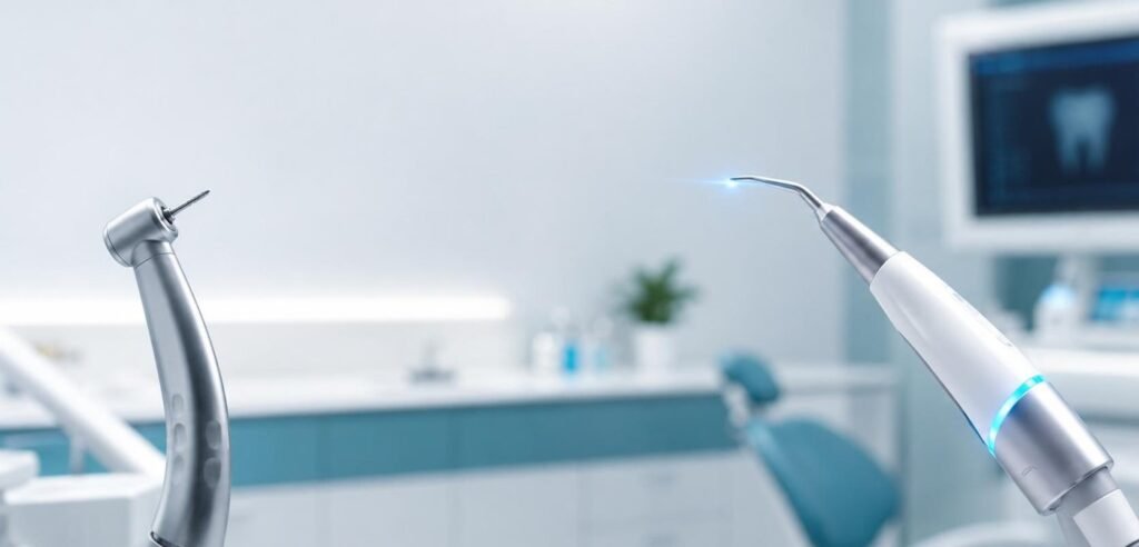
Traditional mechanical cavity preparation based on geometric extension is biological mutilation masquerading as clinical prevention. G.V. Black formulated the extension for prevention doctrine in an era lacking adhesive chemistry, magnification, and biophotonic instruments. Cutting away pristine enamel and healthy peripheral dentin to create mechanical undercut retention locks the tooth into an inevitable cycle of structural failure. That century-old engineering mindset destroys natural teeth. Modern clinical dentistry must prioritize biological preservation above historical procedural habit.
The death spiral of the restored tooth begins with the first aggressive bur cut. An operator cuts an extensive Class II cavity for an incipient proximal lesion. Healthy marginal ridges disappear. Cusp flexure doubles under occlusal loading. Ten years later, recurrent micro-leakage demands a wider composite or amalgam restoration. Cusp fracture follows. The tooth receives a full-coverage crown, which strips away 70% of remaining coronal tooth structure. Pulpal necrosis occurs in 15% of crowned teeth, leading to root canal therapy, post placement, root fracture, and terminal extraction.
Biomechanical Strain and Cusp Deflection: The Physics of Cavity Geometry
Cavity geometry dictates tooth longevity.
Finite element analysis confirms that intact natural teeth function as unified prestressed biomechanical domes. Occlusal masticatory forces transfer through enamel rods into the resilient dentin core, distributing stress evenly down the root sheath into the periodontal ligament. When a high-speed diamond bur transects the marginal ridge to establish a conventional G.V. Black proximal box, cuspal stiffness plummets by 63%. Preparing both mesial and distal marginal ridges in a standard mesio-occlusal-distal configuration reduces overall tooth rigidity by more than 85%.
Sharp internal line angles act as mechanical stress concentrators. Mechanical rotary burs naturally leave 90-degree internal transitions. Under cyclic masticatory loading of 250 Newtons, these geometric corners generate intense microtensile stress peaks that radiate outward toward the cervical margin. Microcracks propagate through sound dentin over millions of chewing cycles. Complete coronal cusp fractures inevitably result.
The Cellular Biology of Carious Dentin: Infected Versus Affected
Dentin is not a uniform block of dead mineral. It is a living, cellular tissue complex traversed by millions of cytoplasmic odontoblastic processes connected directly to the dental pulp chamber.
Carious dentin divides into two distinct histological layers. Outer carious dentin is necrotic and infected. It is irreversibly demineralized, saturated with high bacterial loads, and its collagen scaffolding is completely denatured. It cannot remineralize; it must be excised.
Inner carious dentin is affected but biologically viable. Bacteria are largely absent. While partially demineralized, the underlying cross-linked type I collagen matrix remains structurally intact. If protected from active micro-leakage and sealed beneath a biomimetic restoration, affected dentin remineralizes through mineral ion uptake from pulp and adhesive glass ionomer materials.
Steel burs cannot distinguish between infected and affected layers. Rotating carbide burs rip through soft affected dentin blindly. The clinician, fearful of leaving caries behind, excavates down to hard, sound dentin near the pulp chamber. A clean, pink pinpoint exposure appears. A vital pulp is needlessly violated. What could have been a conservative indirect pulp cap turns into an invasive root canal intervention.
Selective Laser Ablation: The Physics of Differential Absorption
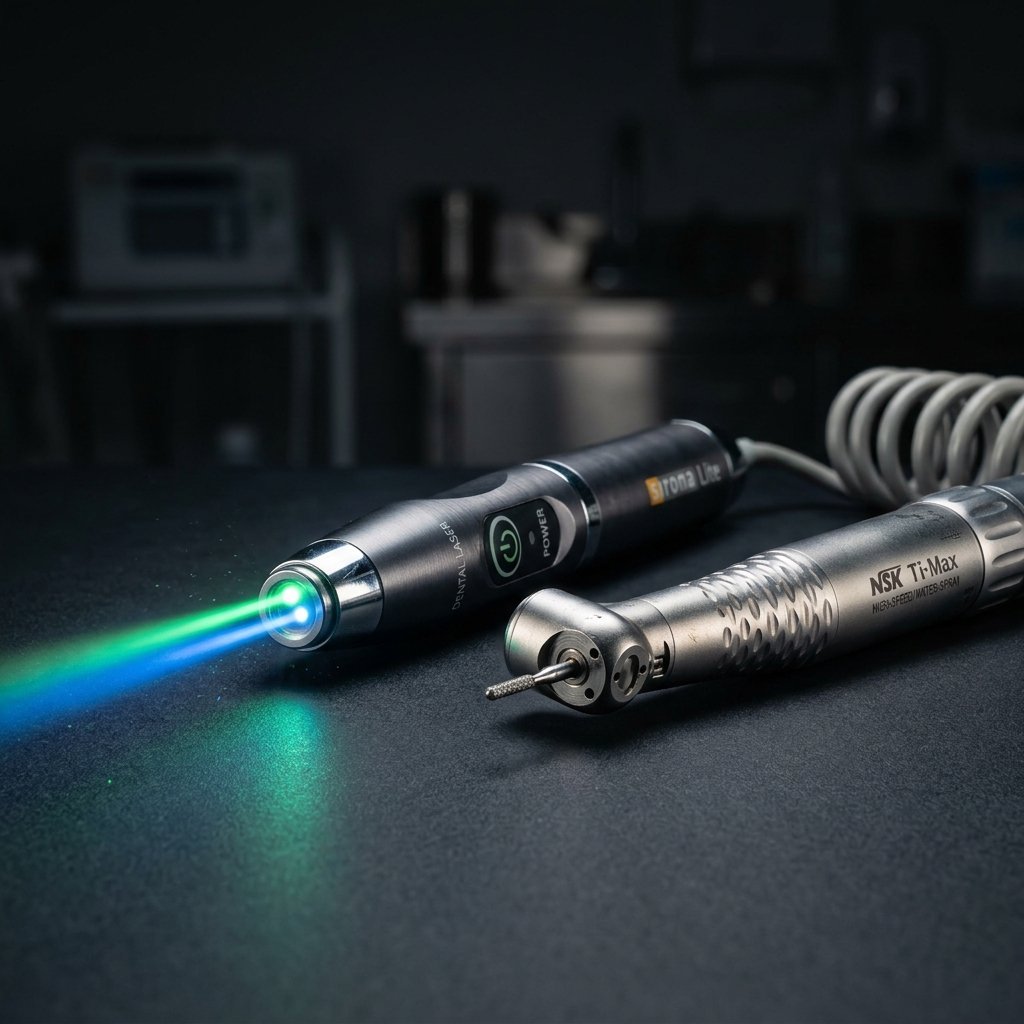
Erbium lasers ablate carious tissue selectively through physical absorption physics.
Water content dictates ablation thresholds. The Er:YAG laser emits photons at 2,940nm, matching the primary absorption peak of liquid water where the linear absorption coefficient reaches an extraordinary 10,000 cm⁻¹. The Er,Cr:YSGG wavelength at 2,780nm exhibits comparable high affinity for bound hydroxyapatite water. Healthy dental enamel contains only 4% water by volume. Sound coronal dentin contains approximately 10% water. Soft, infected carious dentin contains over 25% water.
Carious dentin vaporizes at lower energy densities than healthy tooth structure. When calibrated Er:YAG laser pulses strike a mixed carious lesion, water within the degraded infected layer absorbs photons instantly. Micro-explosions shatter and eject the necrotic tissue. The moment the laser pulse encounters dry, sound dentin with low water content, the cutting speed drops dramatically. The laser functions as an optical tissue selector, stopping automatically before healthy dentinal tubules and pulp horns are compromised.
Co-axial water delivery protects pulpal vitality. Continuous micro-droplet irrigation cools the target site, preventing thermal elevation of the pulp chamber above the critical 5.5°C threshold identified by Zach and Cohen as the trigger for irreversible thermal pulpitis.
No smear layer, no micro-fractures. Mechanical burs spin at 400,000 RPM, producing microscopic crazing lines and a thick smear layer of compressed collagen debris that plugs dentinal tubule openings. Er:YAG laser ablation leaves an open, micro-retentive surface completely free of smear layer. Tubules stand open and pristine, ready for intimate hybridization with modern hydrophilic resin monomers.
Biomimetic Principles: Replicating Natural Tooth Mechanics
Biomimetic dentistry replaces lost tooth structure with synthetic materials that mimic the physical properties of natural dentin and enamel.
Enamel is stiff; dentin is flexible. Dental enamel possesses a high modulus of elasticity (approximately 80 Gigapascals), designed to resist abrasive masticatory wear. Dentin has a lower elastic modulus (approximately 15 Gigapascals), acting as a shock-absorbing stress cushion that protects the tooth from catastrophic splitting forces.
Immediate Dentin Sealing (IDS) preserves the hybrid layer. Sealing freshly cut dentin with a high-performance dentin bonding agent immediately after cavity preparation shields the exposed pulp from bacterial contamination and fluid shifts during provisionalization. Bond strengths increase from 11.5 Megapascals under delayed protocols to over 45 Megapascals with IDS. Polymerization shrinkage occurs without external functional stress.
Deep margin elevation resolves subgingival boundaries conservatively. Rather than subjecting the patient to aggressive surgical crown lengthening that strips away crestal bone support, the operator isolates the subgingival margin under rubber dam and elevates the margin coronally with a micro-hybrid composite. The biological width remains unviolated.
Fiber-reinforced composites eliminate catastrophic fractures. Placing woven polyethylene fiber ribbons beneath deep composite restorations redistributes occlusal stresses horizontally, dissipating tensile forces and preventing vertical root fractures. The tooth flexes naturally as a unified biological unit.
Ethical Responsibility in Restorative Practice
Tooth structure is a non-renewable biological resource. Enamel cannot regenerate once cut away with a high-speed diamond bur. Every millimeter of natural biological tissue preserved in an eighteen-year-old patient represents thirty additional years of functional masticatory health.
Dentists must reject aggressive prep models. Transitioning from aggressive full-coverage crowns to conservative partial-coverage onlays, inlays, and ultra-conservative laser preparations is an ethical obligation. We must view healthy tooth structure with the reverence a vascular surgeon grants to human blood vessels.
Minimally invasive dentistry is not an aesthetic trend; it is clinical bio-engineering at its highest level. Respecting biological boundaries ensures patient longevity.
Controlled retrospective clinical data tracked across international biomimetic research centers shows that teeth restored using selective caries excavation, immediate dentin sealing, and partial-coverage bonded onlays maintain a 97.4% pulp vitality survival rate at ten years. In contrast, teeth receiving traditional full-coverage crowns suffer irreversible pulpal necrosis requiring endodontic intervention in 16.8% of cases over the identical observation period.
Leave a Reply