
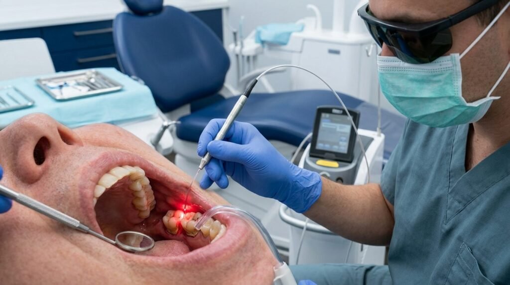
Subgingival bacterial biofilms drive chronic inflammatory destruction within the periodontal pocket. Ultrasonic debridement and curettes often fail to remove pathogens deep in dentinal tubules. Periodontal pathogens such as Porphyromonas gingivalis, Treponema denticola, and Tannerella forsythia possess virulence factors that invade soft tissue pocket walls. Consequently, adjunctive laser technologies have emerged to enhance microbial reduction and modulate host inflammatory responses. Wavelength specificity dictates the photothermal interactions governing tissue ablation and bacterial kill rates. Both Nd:YAG (1064 nm) and diode lasers (810-980 nm) target endogenous chromophores including hemoglobin and pigmented proteins. Understanding their physical and optical characteristics remains vital for achieving predictable clinical attachment gains without thermal collateral damage.
Optical Physics and Chromophore Absorption in the Periodontal Pocket
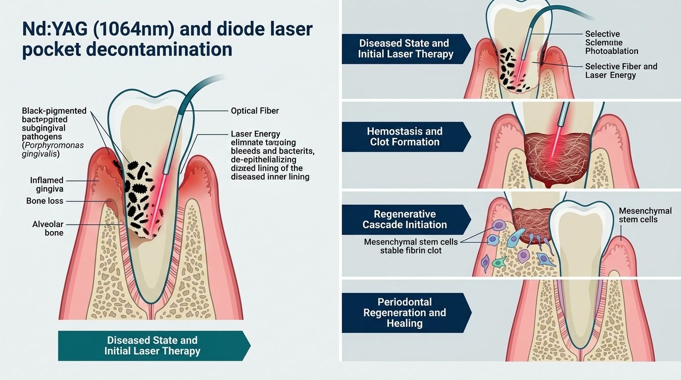
Laser energy delivered through flexible optical fibers penetrates deep into compromised sulcular environments. The 1064 nm Nd:YAG wavelength exhibits low absorption in pure water and hydroxyapatite but shows selective affinity for melanin, black-pigmented bacteria, and oxyhemoglobin. Because red complex anaerobes incorporate hemin into their outer membranes, Nd:YAG radiation achieves selective photoablation of these micro-organisms. Pigmented bacterial walls absorb light energy rapidly, leading to instantaneous thermal lysis and structural protein denaturation. Similarly, diode lasers operating across the 810 to 980 nm spectrum exhibit strong absorption in hemoglobin and melanin. These characteristics enable targeted removal of diseased sulcular epithelium while preserving healthy connective tissue and cementum. Light penetration extends several hundred microns beyond the physical reach of curette tips, neutralizing pathogens that colonize lateral pocket walls.
Energy delivery requires careful calibration of pulse duration, repetition rate, and tip movement. Continuous wave diode delivery can cause unintended thermal accumulation within thin alveolar bone plates. Gated or pulsed emission modes provide essential thermal relaxation times, preventing destructive pulp or periodontal ligament necrosis. Fiber optic diameters between 300 and 400 microns facilitate parallel access to deep narrow defects. Practitioners must maintain continuous, sweeping fiber movement from apical to coronal positions. Halting the laser fiber tip against root surfaces risks cementum melting, root cracking, or focal ankylosis.
The LANAP Protocol and Cementum-Mediated New Attachment
The Laser-Assisted New Attachment Procedure (LANAP) utilizes pulsed free-running Nd:YAG energy under a standardized clinical framework. Histological evidence demonstrates true periodontal regeneration following specific LANAP sequences, including new cementum, periodontal ligament fibers, and functional alveolar bone. The protocol begins with shallow sulcular laser passes using short microsecond pulse widths. This initial exposure disrupts bacterial cell walls, vaporizes granulomatous tissue, and loosens tenacious subgingival calculus without damaging mineralized root substrates. Subsequent piezoceramic debridement removes denatured mineral concretions from the contaminated root surface. Then, a secondary deeper laser pass at modified pulse characteristics initiates blood coagulation, creating an autologous stable fibrin clot.
This stable thermal fibrin clot acts as a biological barrier against rapid epithelial downgrowth. Long junctional epithelium formation represents the default healing pattern following conventional mechanical scaling. By sealing the sulcular entrance with a dense hemocoagulum, the Nd:YAG laser provides slow-migrating multipotent mesenchymal cells access to denuded root surfaces. Fibroblastic proliferation occurs across an organized fibrin matrix rich in crosslinked platelet factors. Animal and human biopsies confirm coronal progression of functional connective tissue attachment without foreign body reactions. Maintaining clot integrity during the immediate postoperative window proves decisive; probing depths must not be measured for several months.
Bactericidal Kinetics Against Red Complex Micro-Organisms
Microbiological monitoring reveals substantial reductions in anaerobic microbial loads following targeted near-infrared laser therapy. Porphyromonas gingivalis produces black-pigmented iron protoporphyrins that render it exceptionally vulnerable to 810 to 1064 nm photothermal disruption. Studies using real-time polymerase chain reaction demonstrate multi-log reductions in subgingival bacterial loads within minutes of irradiation. Unlike systemic antibiotics, laser energy induces physical rupture of bacterial membranes without generating pharmacologic resistance mutations. Lipopolysaccharide endotoxins embedded within diseased root cementum are rendered biologically inert through thermal degradation. This detoxification creates an architecturally receptive root substrate suitable for fibroblast reattachment and hemidesmosome assembly.
Nevertheless, bacterial recolonization begins if supragingival plaque control remains compromised. Lasers alter the microbial ecology temporarily, establishing an ecological window favoring non-pathogenic commensal colonization. Combining laser debridement with strict maintenance recalls maximizes suppression of dysbiotic microbial consortia. Clinicians must realize that subgingival phototherapy complements, but does not entirely replace, mechanical instrumentation. Ultrasonic debridement remains necessary to disrupt hard calculus matrices that reflect rather than absorb near-infrared photonic streams.
Thermal Safety Thresholds and Periodontal Tissue Biology
Biological tissues display variable tolerance to laser-induced thermal elevation. A temperature increase exceeding 5 degrees Celsius inside the dental pulp initiates pulpal hyperaemia, while an increase exceeding 10 degrees Celsius causes irreversible necrosis and periodontal ligament thrombosis. To maintain physiological safety, average laser power should rarely exceed 1.0 to 1.5 Watts in closed periodontal environments. Water cooling, pulse pauses, and intermittent suction evacuate trapped heat effectively. Thin biotypes require lower energy fluences compared to thick fibrotic gingival margins.
Histological studies on diode lasers indicate that superficial carbonization layers must be avoided. Charring creates an opaque black barrier that prevents forward light penetration, trapping heat at the fiber tip. When carbonization occurs, surrounding connective tissue suffers microvascular thrombosis and delayed collagen synthesis. Proper fiber cleaving, stripping, and contact management eliminate debris build-up at the tip. Fiber tips should be cleaned repeatedly with moist sterile gauze during deep pocket debridement.
Clinical Outcomes and Methodological Evidence
Systematic reviews evaluating laser pocket decontamination demonstrate modest but statistically significant benefits in probing depth reduction and clinical attachment level gains. Deep periodontal pockets exhibiting baseline depths above 6 millimeters derive the greatest measurable advantage from adjunctive laser irradiation. Furcation involvements, which resist conventional hand instruments due to anatomical complexity, exhibit reduced bleeding on probing following fiber optic decontamination. Longitudinal studies tracking tooth retention demonstrate reduced surgical intervention requirements when laser therapy integrates into active periodontal management programs. Patient-reported outcome measures reflect lower intraoperative discomfort, reduced postoperative analgesic consumption, and minimal dentinal hypersensitivity.
Heterogeneity across published clinical trials stems from variable power settings, fiber sizes, and lack of standardized reporting. Scientific progress depends on strict adherence to clear dosimetry reporting, including spot size, peak power, repetition frequency, and energy density. Wavelength selection must match clinical pathology: Nd:YAG excels at deep selective soft-tissue curettage and clot induction, whereas diode wavelengths provide accessible, cost-effective bacterial decontamination and gentle biostimulation. Practitioners must combine rigorous physical knowledge with patient-specific anatomy to achieve durable clinical outcomes.
References
- Yukna, R. A., Carr, R. L., & Evans, G. H. (2007). Histologic evaluation of an Nd:YAG laser-assisted new attachment procedure in humans. The International Journal of Periodontics & Restorative Dentistry, 27(6), 577-587.
- Cobb, C. M. (2017). Lasers and the treatment of periodontitis: the essence and the noise. Periodontology 2000, 75(1), 205-295.
- Qadri, T., Miranda, L., Tunér, J., & Gustafsson, A. (2005). The short-term effects of low-level lasers as adjunct to conventional therapy in the treatment of periodontal inflammation. Journal of Clinical Periodontology, 32(7), 714-719.
- Aoki, A., Mizutani, K., Schwarz, F., Sculean, A., Yukna, R. A., Ghanbari, B. K., … & Ishikawa, I. (2015). Periodontal and peri-implant wound healing following laser therapy. Periodontology 2000, 68(1), 217-269.
- Miron, R. J., & Sculean, A. (2021). Biomaterials and regenerative strategies in periodontology. Clinical Oral Investigations, 25(6), 3365-3375.
Leave a Reply