
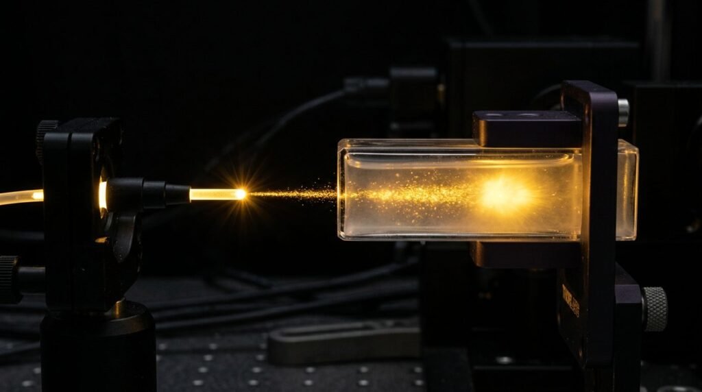
Surgical and non-surgical management of advanced periodontitis demands the eradication of dysbiotic subgingival biofilms and the establishment of a biocompatible root surface capable of supporting stable periodontal reattachment. Mechanical scaling and root planing (SRP) utilizing mechanical curettes and ultrasonic tips effectively debulks calcified calculus but routinely produces an amorphous smear layer packed with bacterial endotoxins, while simultaneously removing extensive layers of healthy root cementum. Excessive cementum removal exposes permeable dentinal tubules, creating postoperative sensitivity and hindering new connective tissue attachment. Application of the pulsed neodymium-doped yttrium aluminum garnet (Nd:YAG) laser emitting at 1064 nm introduces a selective photothermal protocol that vaporizes dark-pigmented periodontal pathogens, removes diseased pocket epithelium, and preserves native root cementum to facilitate durable fibrin clot stabilization.
The biophysical selectivity of the 1064 nm wavelength is governed by its preferential absorption in melanin, exogenous pigments, and black-pigmented anaerobes, accompanied by near-zero absorption in hydroxyapatite and water. Major periopathogens implicated in destructive periodontitis, including Porphyromonas gingivalis, Tannerella forsythia, and Prevotella intermedia, synthesize surface-bound hemin and protoporphyrin IX pigments that act as concentrated endogenous chromophores. When the pulsed Nd:YAG laser beam penetrates the periodontal pocket, photonic energy is absorbed selectively by these chromophore-rich bacterial colonies. This localized optical absorption causes instantaneous photothermal lysis of bacterial membranes at energy fluences far below the thermal threshold of adjacent root dentin. Concurrently, bacterial lipopolysaccharides (LPS) entrenched within the outer cementum matrix undergo thermal inactivation and molecular denaturation, converting a cytotoxic root surface into a biocompatible biological substrate.
Selective ablation of diseased pocket epithelium represents a distinguishing characteristic of the 1064 nm pulsed protocol. Conventional open-flap surgery requires invasive scalpel incisions and periosteal reflection to excise inflamed granulomatous pocket linings. Under calibrated Nd:YAG parameters, the flexible bare quartz optical fiber (typically three hundred to four hundred micrometers in diameter) is directed toward the soft tissue wall of the pocket, moving parallel to the root axis in a coronal-to-apical sweep. Because hyperemic, chronically inflamed pocket lining contains dense microcapillaries packed with deoxyhemoglobin, the 1064 nm beam selectively ablates the ulcerated pocket epithelium and inflamed lamina propria without damaging underlying healthy alveolar connective tissues. Histological analyses confirm complete de-epithelialization of the pocket wall while retaining deep collagen bundles that serve as scaffolds for periodontal regeneration.
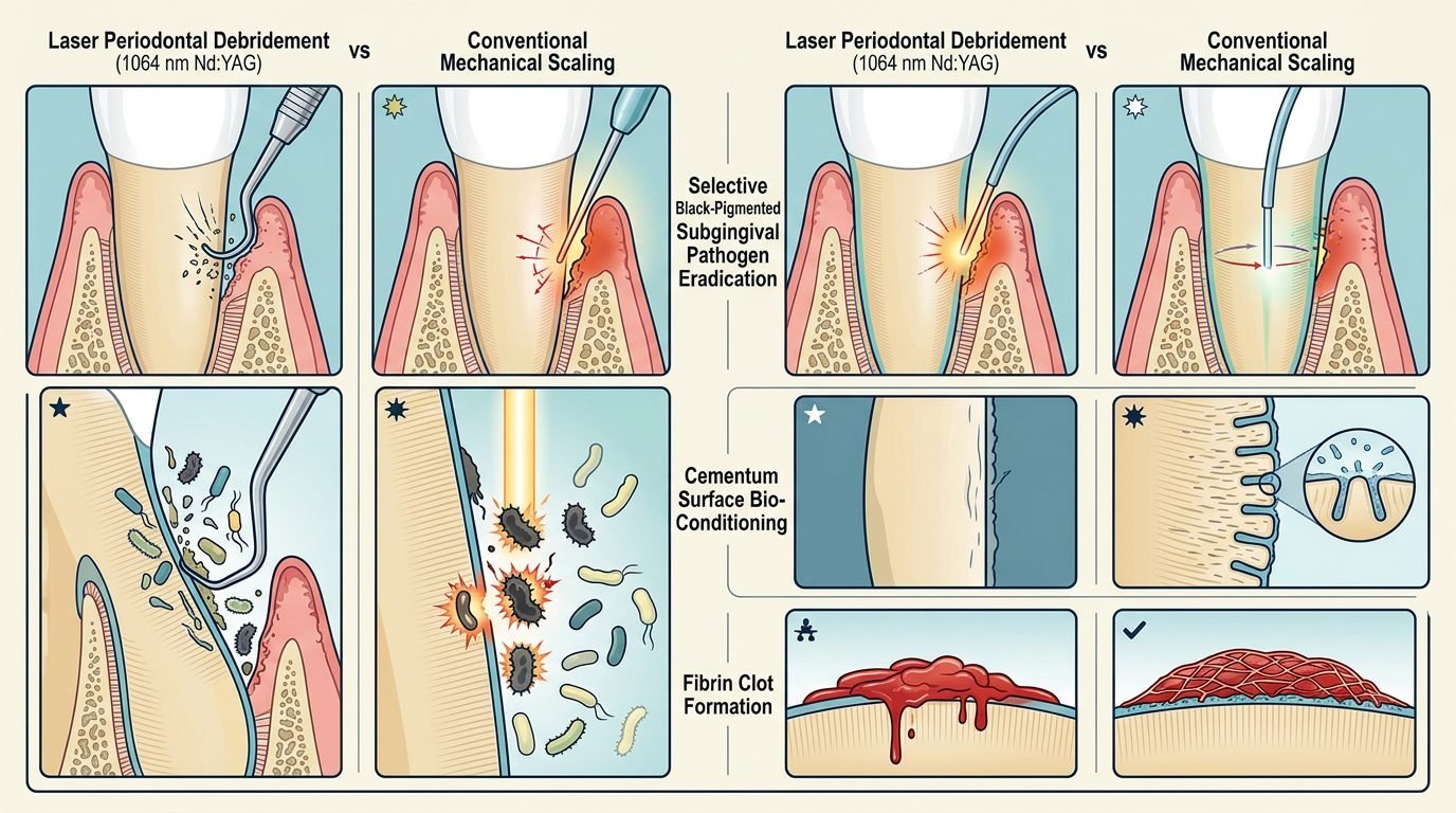
Cementum conditioning and the preservation of fibrous attachment apparatus differ fundamentally between laser and mechanical debridement. Mechanical instrumentation with sharp steel curettes strips away five to thirty micrometers of root cementum per stroke, producing smooth, devitalized root surfaces coated in dense mechanical smear layers that inhibit cell spreading. Conversely, non-contact or light-contact Nd:YAG laser irradiation preserves the underlying structural cementum matrix, creating a micro-textured, smear-free root topography. Scanning electron microscopic (SEM) examinations reveal that laser-conditioned cementum surfaces exhibit open collagen fiber stumps and clean cemental micro-lacunae. This conditioned surface exhibits exceptional surface wettability and surface free energy, creating an ideal biological interface for early mesenchymal stem cell migration and direct osteoblastic differentiation.
Fibrin clot stabilization constitutes the definitive physiological requirement for true periodontal regeneration rather than long junctional epithelial repair. If a newly formed blood clot detaches from the instrumented root surface during early wound healing, oral sulcular epithelial cells rapidly migrate apically along the dentin face, establishing an unstable junctional epithelium. A second pass of the Nd:YAG laser at low average power and low pulse frequency is delivered directly into the deep pocket base to generate a stable, cross-linked fibrin thermal seal. The 1064 nm radiation gently heats hemolyzed blood, activating intrinsic coagulation cascades and promoting stable polymerization of high-density fibrin clots that adhere securely to the micro-textured root cementum. This dense biological seal prevents downgrowth of overlying gingival epithelium, isolating the healing space to permit true connective tissue attachment with newly formed Sharpey fibers inserting into regenerated cementum.
Dosimetric parameters must be calibrated rigorously across both distinct surgical passes to avoid collateral thermal necrosis of the alveolar bone or permanent pulpitis. The initial de-epithelialization and bacterial decontamination pass uses a flexible 300-micrometer quartz optical fiber with an average power of 2.0 to 4.0 watts, individual pulse energies calibrated between one hundred and one hundred and fifty millijoules, a pulse repetition rate of twenty to thirty hertz, a pulse width of one hundred to one hundred and fifty microseconds, and energy densities between fifteen and thirty joules per square centimeter. The fiber tip is held in a light contact mode parallel to the root surface, moving continuously in an apical-to-coronal sweeping direction. The second pass, designed specifically to achieve controlled hemostasis and secure the biological fibrin clot, utilizes lower, non-ablative settings: 1.0 to 2.0 watts, twenty hertz, pulse width of one hundred microseconds, and an energy density of five to ten joules per square centimeter. Throughout laser delivery, the optical fiber must remain in continuous, sweeping motion, never remaining stationary for more than one second at any anatomical location. Clinicians must monitor pulpal and bony temperature dynamics, ensuring that cumulative intrapulpal temperature elevation does not exceed the recognized three-to-five-degree-Celsius safety threshold.
Longitudinal clinical trials demonstrate that full-mouth Nd:YAG laser-assisted periodontal regenerative protocols produce significant clinical attachment level (CAL) gains, substantial probing depth (PD) reductions, and increased radiographic alveolar bone heights at twelve to thirty-six months post-treatment. Compared to conventional scaling and root planing alone, patients treated with adjunctive Nd:YAG debridement achieve greater pocket closure in deep infrabony defects (probing depths greater than six millimeters). Human histological biopsy studies substantiate these clinical findings, demonstrating true new cementum formation with inserting collagen fibers and regenerated coronal alveolar bone without signs of long junctional epithelial encapsulation.
Postoperative wound management protocols require total mechanical immobility of the treated sites. Because the success of laser-mediated periodontal regeneration depends on maintaining the primary fibrin clot intact against the root surface, mechanical probing, subgingival instrumentation, and vigorous flossing are strictly contraindicated at treated sites for nine to twelve months post-surgery. Premature mechanical disruption shears developing collagen micro-anchors, converting a regenerating connective tissue complex into an inflamed pocket. Patients must be maintained on chemical plaque control using 0.12 percent chlorhexidine gluconate oral rinses and ultra-soft manual toothbrushes restricted to coronal tooth surfaces during the initial three months of healing.
Regarding clinical practice, incorporating the 1064 nm Nd:YAG laser into comprehensive periodontal therapy bridges the historic gap between conventional mechanical debridement and invasive open-flap surgical resection. By successfully combining targeted photo-destruction of pigmented anaerobes, selective soft-tissue pocket de-epithelialization, root cementum preservation, and cross-linked thermal fibrin clot sealing, pulsed Nd:YAG photonic irradiation creates the precise biological microenvironment necessary for true periodontal regeneration. Clinicians who adhere strictly to verified dosimetric parameters and rigid postoperative tissue immobilization guidelines can achieve predictable connective tissue reattachment, radiographic bone fill, and long-term tooth retention even in severely compromised dentitions.
Leave a Reply