

Needle phobia in the dental operatory represents a neurovascular vulnerability capable of triggering autonomic collapse, bradycardia, and cerebral hypoperfusion during routine local anesthesia. Trypanophobia affects ten percent of the adult population and accounts for over sixty percent of dental medical emergencies. Unlike standard situational anxieties that manifest as sympathetic arousal, needle phobia exhibits a biphasic autonomic reflex: initial tachycardia and hypertension rapidly collapse into parasympathetic vagal stimulation, causing peripheral vasodilation and cerebral ischemia. When clinicians mistake these symptoms for simple nervousness, patients experience syncopal episodes and trauma driving dental avoidance. Preventing these crises requires hemodynamic management, applied tension techniques, computerized micro-dosing systems, and chairside desensitization.
Pathophysiology of the Biphasic Vasovagal Response
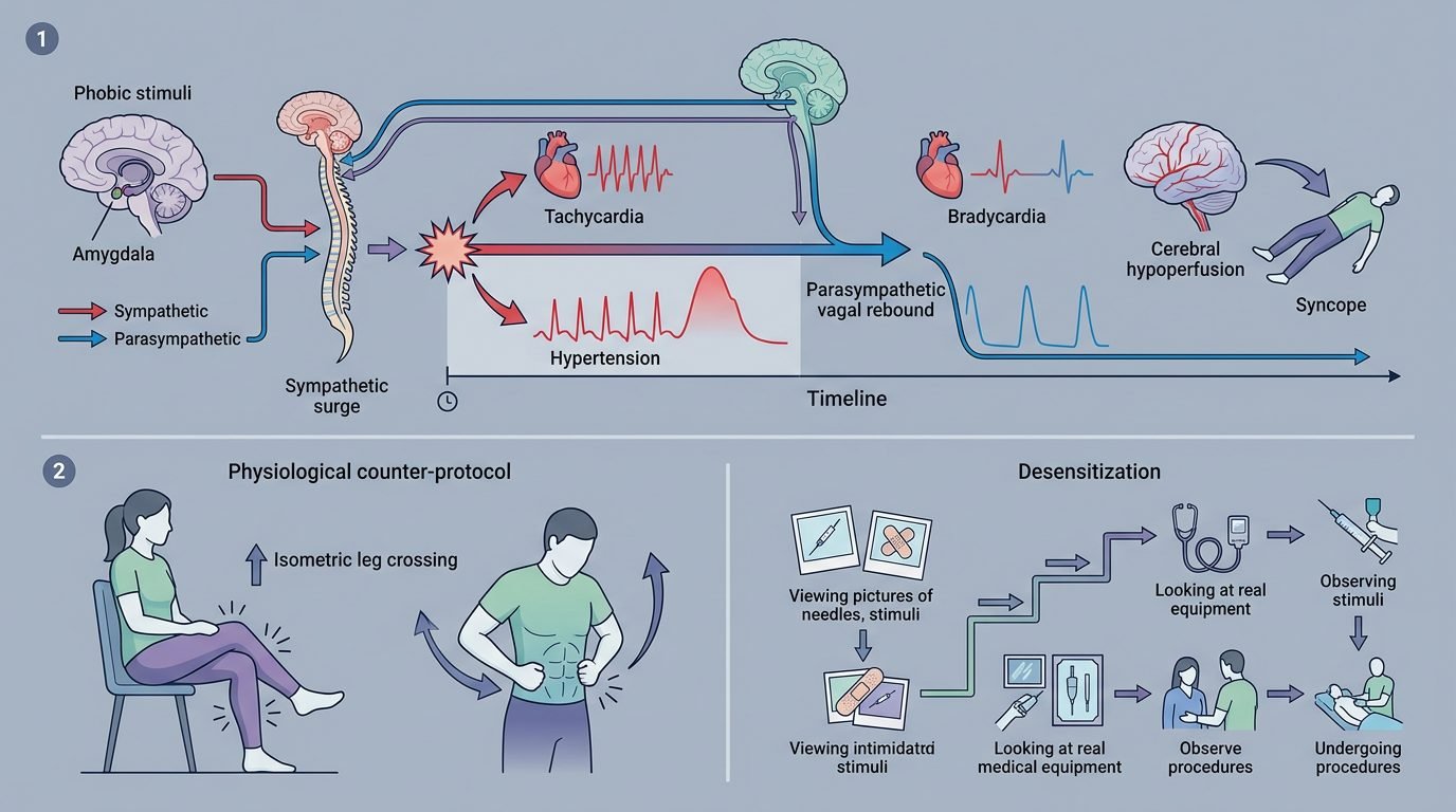
The human vasovagal syncopal reflex represents an evolutionary adaptation mediated through the nucleus tractus solitarius and dorsal vagal motor nucleus in the brainstem. In the dental operatory, exposure to the metal aspirating syringe, auditory cues, or anticipatory fear of mucosal puncture activates the amygdala. The sympathetic nervous system fires during Phase One, elevating serum catecholamines, increasing heart rate, and raising systemic arterial blood pressure.
Within seconds, this sympathetic surge triggers an overcompensating parasympathetic rebound known as Phase Two. Massive vagal efferent discharge to the sinoatrial node induces sinus bradycardia or transient cardiac standstill. Concurrently, sympathetic tone to peripheral arterioles drops sharply, causing widespread pooling of blood in skeletal muscle vascular beds. Mean arterial pressure falls precipitously below fifty millimeters of mercury. Deprived of oxygenated blood, the cerebral cortex fails to sustain consciousness, producing dizziness, diaphoresis, pallor, visual tunneling, and sudden postural collapse.
The Applied Tension Protocol: Counteracting Cerebral Hypoperfusion
Standard psychological relaxation techniques, such as diaphragmatic breathing or progressive muscle relaxation, are dangerous when applied to patients experiencing active vasovagal prodrome. Inducing somatic relaxation during the parasympathetic rebound phase accelerates peripheral vasodilation, precipitating immediate syncope. Clinicians must instead teach patients the Applied Tension Technique developed by Öst.
Applied tension actively raises systemic blood pressure through voluntary isometric muscle contraction. When the patient detects early prodromal symptoms (yawning, nausea, cold sweat, or visual blurring), the clinician instructs them to contract the large skeletal muscles of the legs, buttocks, and abdomen for fifteen to twenty seconds until a warm flush is felt in the face. The patient releases tension partially for twenty seconds, without fully relaxing, repeating the cycle five times. This mechanical pump forces pooled venous blood back into central circulation, maintaining cerebral perfusion pressure above the critical syncopal threshold during injection.
Pharmacological and Technological Innovations: Anesthetic Buffering and Computerized Delivery
The stinging pain associated with conventional local anesthetic infiltration directly amplifies the neurogenic trigger of vasovagal reflex arcs. Commercial dental anesthetic cartridges containing epinephrine are manufactured with an acidic pH between 3.0 and 3.5 to prevent catecholamine oxidation. Injecting an acidic solution into neutral tissue (pH 7.4) causes acute nociceptive firing in unmyelinated C-fibers before tissue buffering permits base liberation and neural blockade.
Chairside anesthetic buffering neutralizes this biological problem. Mixing 8.4 percent sodium bicarbonate with 2 percent lidocaine in a 1:10 volume ratio immediately elevates the solution pH to a physiological 7.35 to 7.45. This biochemical conversion increases the concentration of non-ionized lipid-soluble base molecules by more than one thousand percent, accelerating nerve membrane penetration and eliminating acidic injection pain. Furthermore, computerized local anesthesia delivery systems, such as The Wand, utilize dynamic pressure-sensing technology to deliver micro-droplets at an exact rate of one drop every two seconds, remaining below sensory pain thresholds and neutralizing autonomic fear.
Trauma-Informed Chairside Positioning and Visual Shielding
Physical operatory positioning provides mechanical protection against syncopal loss of consciousness. Treating patients with confirmed trypanophobia in an upright posture invites gravity-dependent cerebral ischemia. The clinician must position the patient in a semi-supine or horizontal posture before dental equipment is assembled.
Visual shielding must be maintained rigorously. Uncapping syringes, testing anesthetic flow, and passing instruments directly over the patient chest trigger immediate sympathetic panic. The dental assistant must prepare syringes entirely outside the patient visual field and transfer instruments behind the headrest below the line of sight. Utilizing opaque protective safety glasses prevents patients from catching reflections of needles in overhead operatory lamps. Clinicians should establish non-judgmental dialogue, asking, “Would you prefer to keep your eyes closed while we complete this step?” to reinforce patient autonomy.
Graduated In Vivo Desensitization Protocols
Overcoming deep-seated needle phobia requires graduated exposure therapy that decouples the stimulus from autonomic panic. In dedicated preparatory sessions without active restorative work, the clinician introduces needle components in progressive tiers. The process begins with holding an uncapped plastic safety sheath, followed by visual inspection of a fine 30-gauge ultra-short needle resting in the clinician hand.
The subsequent tier introduces tactile exploration, where the blunt side of a capped syringe rests against the patient arm or lip while practicing the applied tension technique. Once the patient maintains stable pulse and oxygen saturation metrics without autonomic spikes, topical anesthetic compounding gel (20 percent benzocaine or a eutectic mixture of lidocaine and prilocaine) is placed on dry mucosa for three minutes. The clinician applies firm blunt pressure using a cotton pellet before performing a micro-dose test infiltration. By proceeding only after explicit patient consent at each tier, the operatory becomes an environment of predictable mastery rather than perceived physical assault.
Managing Acute Syncopal Events Without Iatrogenic Shame
If a vasovagal syncopal episode occurs despite preventive measures, the clinical team must respond with structured emergency management. The dental chair is immediately lowered into the full Trendelenburg position (head lower than feet) to utilize gravity for rapid cerebral blood restoration. The airway is cleared, tight neckwear loosened, and supplemental oxygen delivered at six liters per minute via nasal cannula. Vital signs are monitored continuously.
The psychological aftermath of syncope requires clinical tact. Patients regaining consciousness frequently experience intense embarrassment, confusion, and fear that they caused disruption. Clinicians must de-escalate this emotional crisis through reassuring language: “Your body experienced a normal biological reflex that lowered your blood.
Clinical Synthesis for Empathetic Dental Teams
Managing needle phobia and vasovagal syncope requires dental practitioners to unite vascular physiology with empathetic psychological accommodations. By substituting passive relaxation with active muscle tension, neutralizing injection pain through anesthetic buffering, and honoring patient autonomy with graduated desensitization, clinicians dismantle the physiological mechanisms of syncopal collapse. Transforming the operatory into a sanctuary of hemodynamic safety preserves natural teeth and restores human dignity to patients who have lived in terror of the dental needle.
References
- Hamilton, J. G. (1995). Needle phobia: a neglected diagnosis. Journal of Family Practice, 41(2), 169-175.
- Malamed, S. F. (2015). Medical Emergencies in the Dental Office (7th ed.). Elsevier.
- Öst, L. G., et al. (1984). Applied tension, applied relaxation, and tension-only in the treatment of blood phobia. Behaviour Research and Therapy, 22(3), 249-260.
- Rayman, S., et al. (2010). The Wand: a computerized approach to dental anesthesia for anxious patients. Journal of the American Dental Association, 141(6), 701-705.
- Saatchi, M., et al. (2015). The efficacy of buffered local anesthetics in reducing pain on injection: a systematic review. Journal of Endodontics, 41(10), 1648-1654.
Leave a Reply