

Dental needle phobia constitutes an intense psychiatric and autonomic disturbance that drives chronic avoidance of oral healthcare services. Unlike common situational anxieties that produce steady sympathetic arousal, blood-injury-injection phobias elicit a unique biphasic cardiovascular reflex. The initial anticipatory phase features acute sympathetic activation, characterized by transient tachycardia and peripheral vasoconstriction. However, upon exposure to visual or tactile needle stimuli, the central nervous system undergoes an abrupt parasympathetic surge. Excessive vagal efferent discharge triggers profound cardioinhibitory bradycardia and peripheral vasodilation, resulting in precipitous arterial hypotension and cerebral hypoperfusion. Patients lose consciousness in the dental chair, experiencing acute vasovagal syncope. Conventional verbal reassurance proves ineffective against this hardwired autonomic reflex. Managing trypanophobia requires structured neurobiological intervention, ergonomic positioning, sensory gating, and applied muscle tension techniques that physically prevent cerebral ischemia during dental local anesthetic administration.
The Biphasic Neurocardiogenic Mechanism and the Bezold-Jarisch Reflex
The neurobiology of vasovagal syncope involves complex reciprocal signaling between the medullary solitary tract, the hypothalamus, and the autonomic motor nuclei. During initial visual contact with the injection syringe, the basolateral amygdala activates the locus coeruleus and sympathetic preganglionic neurons. This brief hyperadrenergic phase represents the classic fight-or-flight survival response common to generalized mammalian fear states.
The catastrophic collapse occurs when intense intraventricular myocardial contractions stimulate unmyelinated mechanoreceptive C-fibers located within the inferoposterior ventricular myocardium. In hypovolemic or hyperadrenergic states, the underfilled ventricles contract vigorously around an empty chamber, triggering the Bezold-Jarisch reflex. Mechanoreceptors transmit excessive afferent discharge via the vagus nerve directly into the nucleus tractus solitarii. The brainstem misinterprets this mechanical deformation as extreme systemic hypertension. In response, central descending pathways instantaneously suppress sympathetic vasoconstrictor tone and fire intense cholinergic vagal bursts toward the sinoatrial and atrioventricular nodes.
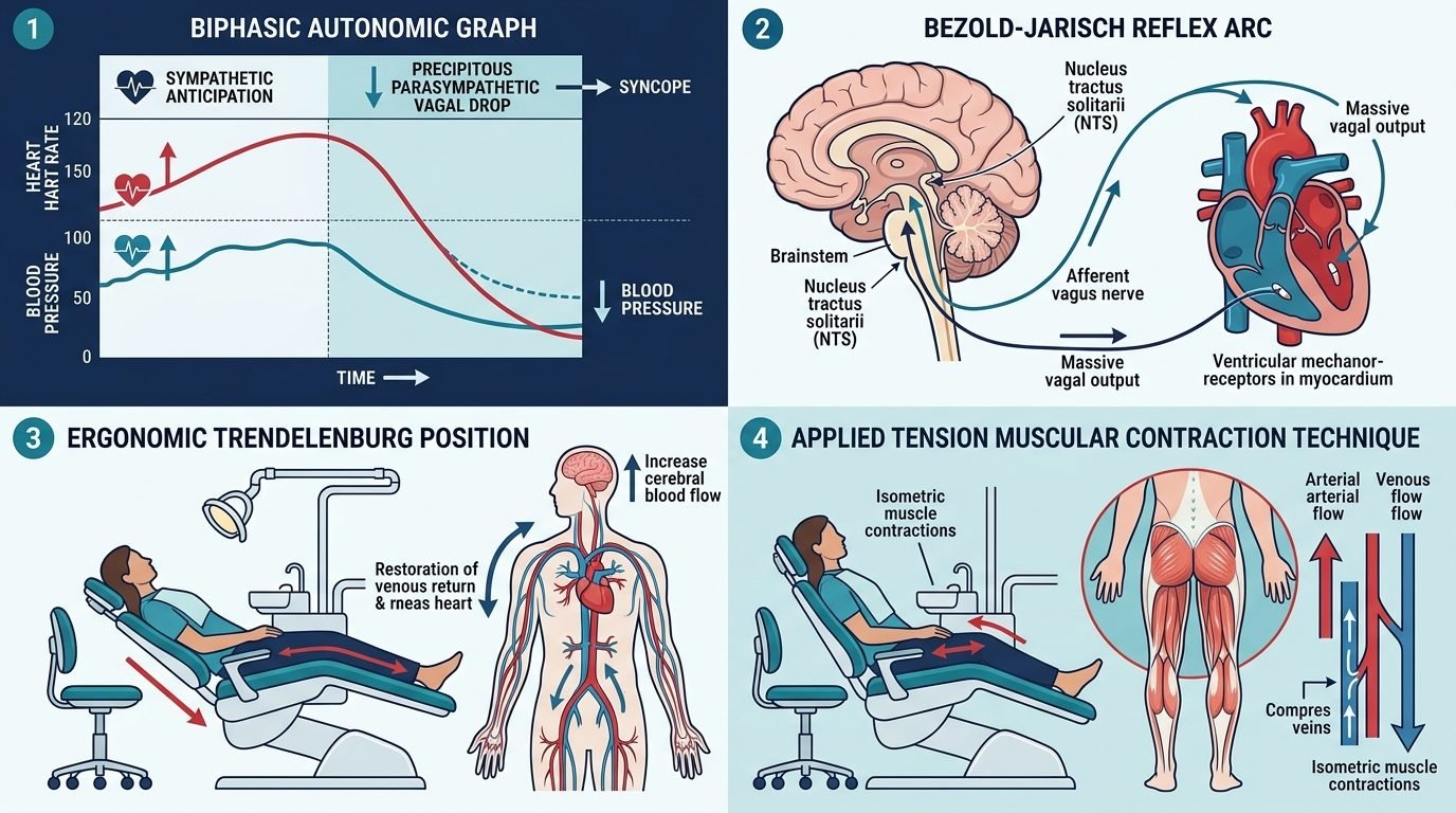
Sinoatrial node firing rates drop precipitously, often leading to sinus bradycardia below forty beats per minute or transient asystolic pauses. Concurrently, systemic vascular resistance collapses as skeletal muscle arterioles dilate. Gravity pools blood in the splanchnic circulation and lower extremities, drastically diminishing venous return to the heart. Cerebral perfusion pressure falls below the critical threshold of thirty millimeters of mercury, depriving cortical and hippocampal pyramidal neurons of oxygen and glucose. Transient loss of consciousness, postural collapse, and brief myoclonic jerking ensue within seconds.
Clinical Manifestations and Prodromal Warning Signs
Recognizing the pre-syncopal prodrome allows the dental team to intervene before loss of consciousness occurs. The transition from sympathetic hyperarousal to parasympathetic collapse takes between twenty and sixty seconds. The patient exhibits pronounced cutaneous pallor resulting from cutaneous vasoconstriction and sudden blood pooling in deep visceral beds. Profuse diaphoresis develops as cholinergic sympathetic fibers stimulate eccrine sweat glands across the forehead and upper lip.
Patients report sudden epigastric nausea, lightheadedness, auditory dulling, and tunnel vision caused by retinal ischemia. Pupillary dilation shifts to sluggish constriction, and the patient may yawn repeatedly due to brainstem hypoxia. Speech becomes dysarthric or entirely absent as cerebral hypoperfusion deepens. Operating clinicians must continuously observe the patient ocular tracking, facial skin color, and breathing patterns. Proceeding with the injection despite obvious prodromal pallor invites full loss of consciousness, prolonged recovery periods, and severe psychological distress.
Ergonomic Positioning and Hemodynamic Stabilization
Ergonomic intervention forms the first biological defense against neurocardiogenic collapse. Administering intraoral local anesthesia to an upright or semi-reclined needle-phobic patient is clinically hazardous. Placing the patient into a horizontal supine position immediately restores venous return, eliminating the hydrostatic pressure gradient opposing cerebral arterial inflow. When syncopal prodromes appear, the dental chair must be positioned into a mild Trendelenburg tilt, elevating the lower extremities ten to fifteen degrees above the head.
Elevating the legs transfers approximately three hundred to five hundred milliliters of pooled blood from the peripheral venous reservoirs back into the central circulation. Left ventricular end-diastolic volume increases, quenching the Bezold-Jarisch mechanoreceptor firing cascade. The clinician must keep the patient head turned sideways to maintain clear airway patency and prevent tongue prolapse or aspiration of saliva.
Applied Tension: The Physiological Antidote to Vasovagal Collapse
While psychological relaxation techniques benefit ordinary dental anxiety, traditional relaxation is strictly contraindicated during the second phase of trypanophobia. Lowering muscle tone and slowing respiration exacerbates arterial hypotension, accelerating syncope. The evidence-based behavioral intervention of choice is the Applied Tension technique, developed by Öst and validated across international clinical trials.
Applied tension teaches the patient to actively contract major skeletal muscle groups throughout the torso, abdomen, and lower extremities. During the injection, the patient rhythmically tenses their quadriceps, gluteal muscles, calves, and abdominal wall for fifteen to twenty seconds until a warm sensation rises to the head. After a five-second release, contraction cycles repeat continuously until local anesthetic delivery concludes. Mechanical skeletal muscle compression collapses deep intramuscular veins, forcing blood back into the vena cava and raising systemic blood pressure by fifteen to twenty-five millimeters of mercury. This deliberate physical elevation of blood pressure preserves cerebral perfusion, breaking the vasovagal reflex arc completely.
Sensory Distraction, Gate Control, and Chemical Buffering
In conjunction with hemodynamic countermeasures, clinicians must modulate peripheral nociceptive signaling. Applying high-frequency vibration devices adjacent to the mucosal penetration site recruits large-diameter, myelinated A-beta cutaneous fibers. According to Melzack and Wall gate control theory of pain, active A-beta tactile signaling closes the presynaptic substantia gelatinosa gates within the spinal trigeminal nucleus, inhibiting the ascent of small nociceptive A-delta and C-fiber inputs to the thalamus.
Furthermore, standard commercial dental anesthetics exhibit an acidic pH between 3.5 and 4.5, stabilized with sodium bisulfite to prevent epinephrine oxidation. This high acidity stimulates unmyelinated nociceptors, producing intense burning sensations during tissue infiltration. Chairside chemical buffering using 8.4 percent sodium bicarbonate neutralizes the anesthetic solution to a physiological pH of 7.4. Buffered local anesthetics convert ionized molecules into uncharged lipid-soluble bases that cross nerve lipid membranes instantly. Injection pain is virtually extinguished, onset time accelerates, and the visceral shock that activates the Bezold-Jarisch reflex is eliminated.
Conclusion and Integrated Humanized Care Protocols
Overcoming dental trypanophobia demands moving beyond simplistic psychological platitudes toward grounded neurovascular science. Recognizing the unique pathophysiology of the biphasic autonomic reflex allows the modern clinician to safeguard patient hemodynamic stability. Combining the Applied Tension technique, Trendelenburg chair ergonomics, sensory vibratory distraction, and chemical anesthetic buffering empowers needle-phobic patients to undergo necessary dental care without experiencing trauma or syncope. Integrating physiological precision with clinical empathy transforms oral healthcare delivery for the most fearful patient populations.
References
- Öst, L. G., & Sterner, U. (1987). Applied tension, applied relaxation, and sensation focusing in the treatment of blood phobia. Behaviour Research and Therapy, 25(4), 269-282.
- Alboni, P., et al. (2007). The vasovagal syncope as a manifestation of an evolutionary adaptation. Journal of Cardiovascular Medicine, 8(8), 587-593.
- Melzack, R., & Wall, P. D. (1965). Pain mechanisms: a new theory. Science, 150(3699), 971-979.
- Malamed, S. F. (2019). Handbook of Local Anesthesia. Elsevier Health Sciences.
- Armfield, J. M. (2010). Towards a better understanding of dental fear: A review. Australian Dental Journal, 55(1), 2-18.
Leave a Reply