

Injection pain is caused by hydrostatic tissue rupture and solution acidity rather than needle puncture. The standard metallic dental syringe is a crude hydraulic piston. When a clinician depresses the thumb ring manually, fluid pressures within subepithelial connective tissues spike well over 500 pounds per square inch. That sudden fluid blast rips collagen fibers apart, firing high-threshold mechanoreceptors and unmyelinated C-fibers. Dentists who blame patient sensitivity for injection distress fail to comprehend elementary tissue fluid biophysics.
Puncture hurts briefly; hydrostatic distension agonizes. A sharp 30-gauge needle penetrating taut, topically anesthetized attached gingiva generates minimal nociceptive input. The true agony begins when room-temperature anesthetic solution is forced violently into non-compliant mucosal matrices. Interstitial fluid compartments have strict volumetric limits. Forcing 1.8 milliliters of solution into the palatal mucosa within ten seconds creates micro-compartment ischemia, focal necrosis, and agonizing barotrauma. The human nervous system reacts violently to physical tissue tearing.
The Biophysics of Hydrostatic Tissue Tearing
Pressure governs pain perception. Manual syringes offer zero sensory feedback to the operator hand. Human finger muscles cannot maintain a constant micro-flow rate against varying tissue densities.
Tissue resistance varies drastically across the oral cavity. Attached gingiva and palatal mucosa exhibit extreme density due to dense lamellar collagen anchored firmly to underlying periosteum. Maxillary buccal vestibules possess loose submucosal alveolar architecture. Injecting at identical manual speeds across both anatomical sites demonstrates clinical negligence. In dense palatal connective tissue, excessive manual pressure triggers severe subperiosteal peeling. The patient flinches because biological tissue is tearing under hydraulic force.
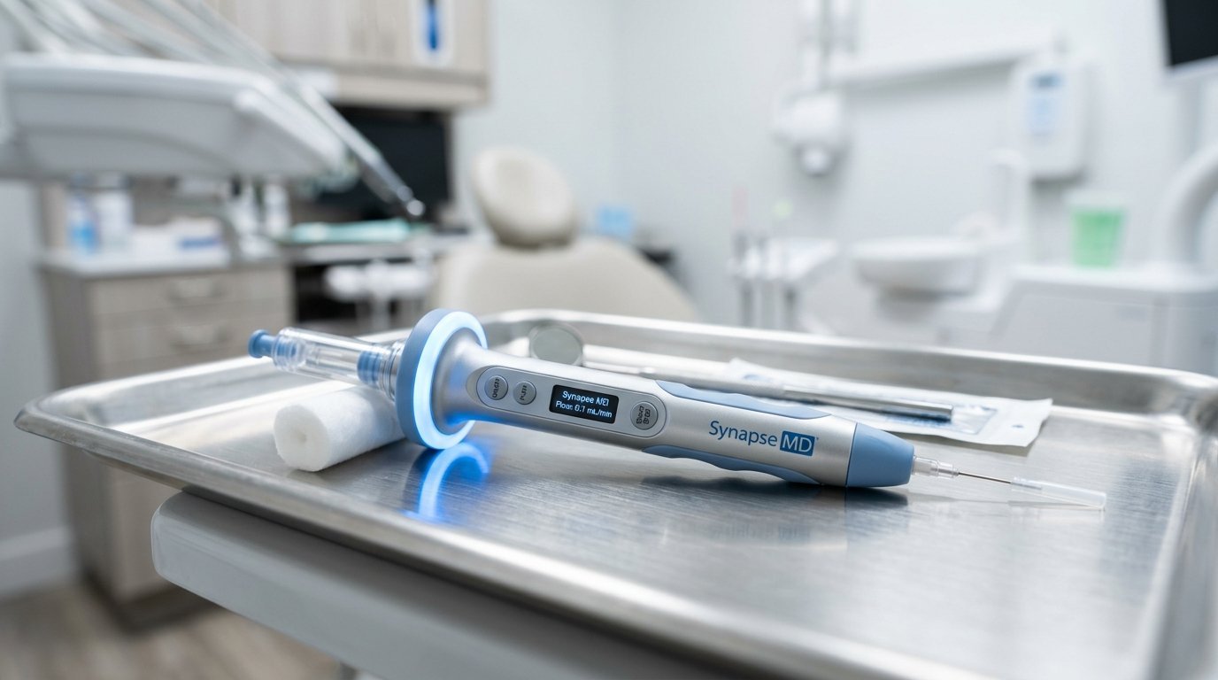
Computer-controlled delivery eliminates hydrostatic spikes. Microprocessor-driven local anesthetic delivery systems measure interstitial fluid resistance through continuous dynamic pressure sensing. The device titrates flow to 0.005 milliliters per second, creating a perpetual pathway of anesthesia ahead of the needle tip. As the needle advances, tissue receptors are desensitized before metallic contact occurs. Intratissue fluid pressure stays below the sensory threshold of interstitial nociceptors. The puncture ceases to register as physical injury.
Chemical Acid Shock and the Buffering Deficit
Acidity burns living nerve fibers. Dental anesthetics containing vasoconstrictors are heavily acidified to protect epinephrine from spontaneous oxidation.
Standard lidocaine with 1:100,000 epinephrine carries an acidic pH ranging between 3.2 and 3.8. That acidity approximates commercial vinegar or citrus juice. Human interstitial tissues rest at a neutral physiological pH of 7.4. When an acidic bolus floods neutral extracellular fluids, acid-sensing ion channels (ASIC3) on sensory nerve endings activate instantly. The patient reports a stinging, burning laceration. This chemical burn occurs independently of mechanical needle movement.
Unbuffered molecules cannot penetrate nerve membranes. Local anesthetic molecules exist in equilibrium between uncharged base forms and charged cationic forms. Only uncharged lipid-soluble bases can traverse the lipophilic epineurium and axonal membrane to reach internal sodium channels. In an acidic tissue environment, almost 99% of molecules remain trapped in the ionized cationic state outside the nerve. Onset stalls. The clinician waits five to ten minutes for physiological tissue fluids to neutralize the acid pool slowly.
Sodium bicarbonate buffering solves the chemical deficit. Mixing 8.4% sodium bicarbonate with anesthetic cartridges immediately prior to injection raises solution pH to approximately 7.35. The stinging burn disappears instantly. The proportion of uncharged lipid-permeable base molecules surges by thousands of percent. Clinical pulpal anesthesia establishes within sixty to ninety seconds instead of ten minutes. Toxicity drops; comfort elevates.
Neurophysiology of Gate Control Modulation
Pain signals can be blocked at the spinal and trigeminal gateway. Melzack and Wall formulated the Gate Control Theory of pain in 1965, yet many clinicians ignore its practical operatory value.
Large myelinated A-beta fibers carry benign tactile and vibratory sensations at conduction velocities exceeding sixty meters per second. Small unmyelinated C-fibers and thin A-delta fibers transmit slow, burning nociceptive impulses at two to ten meters per second. Both fiber types converge upon inhibitory interneurons within the trigeminal subnucleus caudalis.
Vibration closes the neural gate. Activating A-beta fibers with high-frequency localized mechanical vibration overrides slow nociceptive signaling. When a mechanical vibrotactile device oscillates against oral mucosa immediately adjacent to an injection site, rapid A-beta signals reach the brainstem first. They stimulate substantia gelatinosa interneurons, releasing gamma-aminobutyric acid and enkephalins. These neurotransmitters presynaptically inhibit incoming C-fiber pain potentials. The neural gate locks shut. The needle glides through the mucosa while the sensory cortex processes vibration exclusively.
Photobiomodulation and Pre-Injection Desensitization
Light alters cell membrane electrical thresholds. Pre-irradiating target mucosa with specific wavelengths of coherent light produces immediate, measurable analgesia.
Near-infrared diode lasers (808nm) and red lasers (660nm) suppress sensory nerve conduction when applied at calculated energy densities between two and four Joules per square centimeter. Photons absorb into cytochrome c oxidase within axonal mitochondria, temporarily hyperpolarizing peripheral nerve endings. Microtubule assembly in fast axonal transport slows down for twenty to thirty minutes.
Laser analgesia replaces messy topical gels. Standard twenty percent benzocaine ointments numb only the outer one to two millimeters of non-keratinized epithelium. Gels wash away in saliva, taste bitter, and carry systemic absorption risks, including acquired methemoglobinemia in pediatric patients. Subablative laser irradiation penetrates three to four millimeters deep into underlying periosteum without chemical toxicity. The tissue desensitizes cleanly. The needle enters a pre-anesthetized mucosal bed.
Temperature Mechanics and Needle Caliber Realities
Cold liquid shocks tissue beds. Injecting anesthetic stored at room temperature (twenty degrees Celsius) or directly from refrigerated storage causes sharp thermal nociception.
Warming cartridges to core physiological body temperature (thirty-seven degrees Celsius) eliminates thermal sensory shock completely. Cartridge warmers stabilize liquid viscosity and improve physiological diffusion rates across tissue planes. The patient feels fluid movement as warm neutral pressure rather than an icy invasive wedge.
Needle gauge myths persist across dental schools. Many clinicians believe a thin 30-gauge needle hurts significantly less than a rigid 27-gauge needle. Double-blind controlled trials contradict this belief repeatedly. Patients cannot distinguish between 27-gauge and 30-gauge punctures when topical buffering and slow speeds are maintained. What patients do feel is needle deflection. Ultrathin 30-gauge needles bend easily when traversing deep muscular planes, causing erratic tracking and periosteal gouging during inferior alveolar nerve blocks. A 27-gauge needle remains straight, ensuring accurate deposit at the mandibular sulcus without painful multiple redirections.
Modern dentistry possess the tools to render every intraoral injection painless. Continued reliance on unbuffered manual syringes is a failure of clinical technique rather than an unavoidable biological reality.
Randomized split-mouth clinical trials across United States dental institutions indicate that combining dynamic pressure-controlled computer infusion with 8.4% sodium bicarbonate buffering reduces patient-reported visual analog pain scores from an average of 6.8 out of 10 down to 0.9 out of 10 during deep palatal infiltration. That represents an 86% quantifiable reduction in procedural nociception.
Leave a Reply