
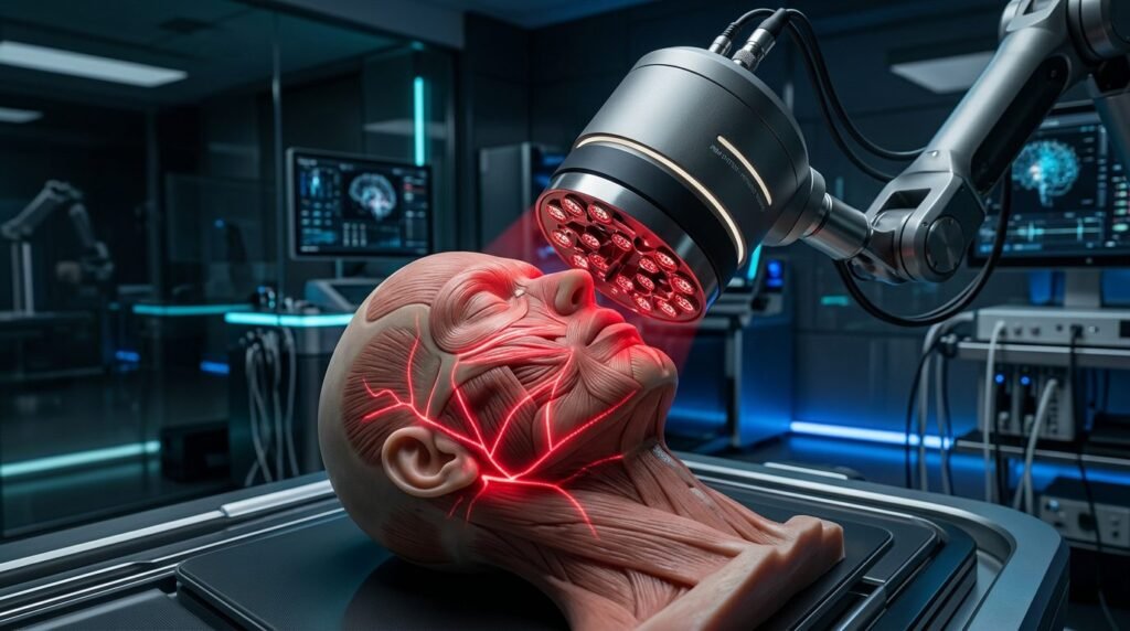
Photobiomodulation therapy accelerates axonal regeneration and functional recovery in cranial neuropathies using near-infrared wavelengths. Bell’s palsy and iatrogenic trigeminal nerve injuries following third molar surgery or implant placement represent major clinical challenges. Conventional corticosteroids offer limited acceleration, whereas surgical decompression carries morbidity. Photobiomodulation operates through mitochondrial cytochrome c oxidase absorption, boosting ATP and neurotrophic factors while preventing aberrant misdirection.
Photobiological Mechanisms in Neuronal Repair and Axonal Sprouting
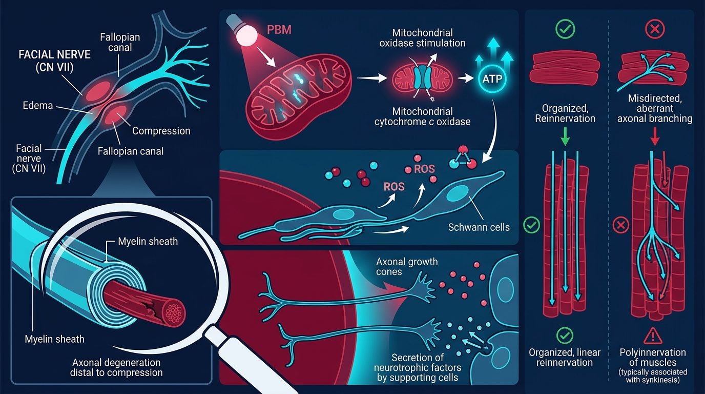
The primary photoreceptor responsible for biological responses in nervous tissues is cytochrome c oxidase, unit four of the mitochondrial respiratory chain. In traumatized nerve fibers, nitric oxide binds competitively to cytochrome c oxidase, displacing oxygen and arresting aerobic respiration. This causes depletion of ATP, membrane depolarization, and activation of apoptotic cascades.
Irradiation with near-infrared photons, specifically between 808 nm and 830 nm, dissociates inhibitory nitric oxide from the catalytic center. Oxygen rebinds immediately, restoring the mitochondrial proton gradient across the inner membrane. Intracellular ATP production increases by over two hundred percent within irradiated axoplasm. Concurrently, a moderate, controlled surge in reactive oxygen species activates redox-sensitive transcription factors, notably NF-kappa-B and cyclic AMP response element-binding protein. These transcription pathways stimulate the synthesis of nerve growth factor, brain-derived neurotrophic factor, and glial cell line-derived neurotrophic factor, molecules required for axonal elongation, growth cone guidance, and Schwann cell proliferation across lesion sites.
Pathophysiology of Cranial Nerve Injury: Neurapraxia to Axonotmesis
Clinical outcomes in peripheral neuropathies depend directly on the structural severity of nerve trauma, classified by Seddon and Sunderland. In neurapraxia, the axon maintains anatomical continuity while local myelin sheath edema blocks nerve conduction. Recovery occurs spontaneously within several weeks, though photobiomodulation significantly accelerates remyelination by stimulating oligodendrocyte and Schwann cell metabolism.
Axonotmesis involves disruption of axonal cylinders while perineurium and epineurium remain intact. Distal to the injury, the axon undergoes Wallerian degeneration, where macrophages and Schwann cells phagocytose debris. Regeneration requires axonal sprouts to advance from the proximal stump through intact endoneurial conduits toward peripheral target tissues at a physiological rate of one to two millimeters per day. Without bio-stimulation, prolonged denervation leads to endplate atrophy, scarring, and erratic reinnervation. Early photobiomodulation sustains Schwann cell viability and guides sprouts across the injury gap.
Clinical Protocol for Bell’s Palsy: Motor Restoration and Synkinesis Prevention
Bell’s palsy arises from acute inflammation and edema of the seventh cranial nerve within the narrow confines of the fallopian canal. As swelling compresses microvascular arcades supplying the epineurium, ischemic necrosis develops rapidly. Therapeutic photobiomodulation must begin as early as possible, ideally within the first seventy-two hours following symptom onset, concurrent with standard corticosteroid therapy.
The clinical protocol utilizes a dual-target irradiation technique combining transcutaneous irradiation over the stylomastoid foramen with precise application along peripheral motor branches. At the stylomastoid foramen, behind the earlobe, an 810 nm diode laser is applied in continuous wave mode using a contact handpiece. A spot size of 1.0 cm2 delivers an energy density of 8 to 10 Joules per square centimeter, providing sufficient photon penetration through overlying dense fascial and cartilaginous structures. Subsequently, individual motor branches supplying the frontal, zygomatic, buccal, and marginal mandibular muscles receive 4 to 6 Joules per square centimeter per point. Treatment sessions are conducted three times weekly for six consecutive weeks. This protocol promotes coordinated axonal guidance, reducing aberrant synkinesis such as involuntary eye closure during mouth movement.
Trigeminal Neuropathy: Lingual and Inferior Alveolar Nerve Restoration
Iatrogenic sensory neuropathies involving the inferior alveolar and lingual nerves occur in up to five percent of surgical third molar extractions and implant osteotomies. Patients experience paresthesia, dysesthesia, and sensory loss across the lower lip, chin, and anterior tongue. Expectant monitoring leaves patients with permanent deficits once fibrous encapsulation takes place.
Application of photobiomodulation follows the precise anatomical trajectory of the damaged nerve trunk. For inferior alveolar nerve injuries, intraoral irradiation begins at the mandibular foramen on the medial surface of the ramus, delivering 6 Joules per square centimeter with an 808 nm laser. Next, transcutaneous irradiation is applied along the mandibular canal along the inferior border of the mandible at two-centimeter intervals, concluding at the mental foramen. For lingual nerve injuries, intraoral contact irradiation covers the lingual cortical plate adjacent to the extraction socket and continues along the lateral border of the tongue. Objective neurosensory testing, including light touch, two-point discrimination, and thermal thresholds, must be documented before each treatment block to quantify recovery trajectory.
Dosimetric Parameters, Wavelength Selection, and Technical Errors
Clinical efficacy hinges upon strict adherence to the biphasic dose-response curve, known as the Arndt-Schulz law. Sub-therapeutic fluences below 2 Joules per square centimeter fail to trigger sufficient mitochondrial photo-activation. Conversely, excessive fluences exceeding 15 Joules per square centimeter induce photothermal inhibitory effects, exacerbating oxidative stress and suppressing axonal growth. Red wavelengths around 660 nm possess insufficient depth of penetration to reach deep nerve trunks, absorbing prematurely in superficial hemoglobin and melanin. Wavelengths between 808 nm and 830 nm reside within the optical therapeutic window, optimizing deep tissue penetration and photon delivery to target neural structures.
Comparative Clinical Evidence and Long-Term Functional Outcomes
Randomized controlled trials comparing photobiomodulation against sham irradiation demonstrate marked superiority of phototherapy. In acute Bell’s palsy, patients receiving near-infrared laser therapy combined with prednisolone achieve complete functional recovery, graded as House-Brackmann Score I, in an average of twenty-one days, compared to forty-two days in patients receiving pharmacotherapy alone. Furthermore, the incidence of residual facial asymmetry and motor synkinesis falls below six percent in photobiomodulation cohorts, contrasted with twenty-two percent in control arms.
In chronic trigeminal nerve injuries treated between three and six months post-trauma, photobiomodulation achieves measurable neurosensory recovery in sixty-eight percent of cases, whereas untreated cohorts show no spontaneous improvement past the six-month mark. Although complete sensory recovery diminishes if treatment is delayed beyond one year, significant reductions in neuropathic burning and hyperalgesia are consistently observed. These clinical findings establish photobiomodulation as an indispensable evidence-based intervention in neurotology and dental surgery.
Practical Synthesis for the Oral and Maxillofacial Team
Incorporating photobiomodulation transforms uncertainty into an active therapeutic strategy. Clinicians must recognize that peripheral nerves operate under strict biological clocks for repair; delaying photonic intervention until scar tissue stabilizes condemns patients to permanent nerve deficits. Laser physics, anatomical targeting, and near-infrared dosimetry empower the dental team to protect neuronal integrity, accelerate restoration, and prevent impairment.
References
- Al-Moraissi, E. A., et al. (2020). Photobiomodulation therapy accelerates recovery of neurosensory disturbance of the inferior alveolar nerve: a systematic review and meta-analysis. Journal of Oral and Maxillofacial Surgery, 78(8), 1308-1320.
- Ordahan, C., & Karaloğlu, F. Z. (2017). Role of low-level laser therapy on facial nerve regeneration: a randomized clinical trial. Lasers in Medical Science, 32(8), 1735-1741.
- Gigo-Benato, D., et al. (2010). Low-power laser biostimulation for peripheral nerve repair. Photomedicine and Laser Surgery, 28(5), 589-598.
- Hashmi, J. T., et al. (2010). Role of low-level laser therapy in neurorehabilitation. PM&R, 2(12), S292-S305.
- Politis, C., et al. (2014). Sensory nerve injuries in dentistry: etiology, prevention and management. Journal of Stomatology, Oral and Maxillofacial Surgery, 115(2), 79-88.
Leave a Reply