
Photobiomodulation therapy relieves intractable intraoral dysesthesia and burning sensations in primary burning mouth syndrome by restoring axonal mitochondrial metabolism and dampening peripheral trigeminal hyperexcitability. Burning mouth syndrome (BMS) is a chronic neuropathic pain condition characterized by continuous, unprovoked burning sensations across clinically normal oral mucosa. Affecting predominantly peri- and postmenopausal women, the condition involves idiopathic neuropathic dysfunction of small unmyelinated and thinly myelinated sensory nerve fibers (A-delta and C-fibers) in the chorda tympani and trigeminal nerve branches. Because physical examination reveals zero visible mucosal ulcerations or erythema, patients face frequent diagnostic dismissal, clinical skepticism, and decades of ineffective empirical therapies. Systemic pharmacological options, such as topical or systemic clonazepam, gabapentin, and tricyclic antidepressants, offer partial symptom suppression while inducing severe drowsiness, ataxia, and cognitive blunting. Photobiomodulation delivers non-thermal red and near-infrared optical energy that re-establishes neurovascular equilibrium, stimulates nerve regeneration, and provides sustained analgesia without systemic toxicity.
Neurobiological Pathophysiology of Burning Mouth Syndrome
Primary burning mouth syndrome represents a distinct neuropathic pain state classified under the International Classification of Orofacial Pain (ICOP). Skin and mucosal biopsy studies employing protein gene product 9.5 (PGP 9.5) immunostaining demonstrate a significant decrease in the density of intraepidermal and subepithelial nerve fibers in the anterior two-thirds of the tongue. This loss of terminal nerve endings reflects a peripheral degenerative small-fiber neuropathy.
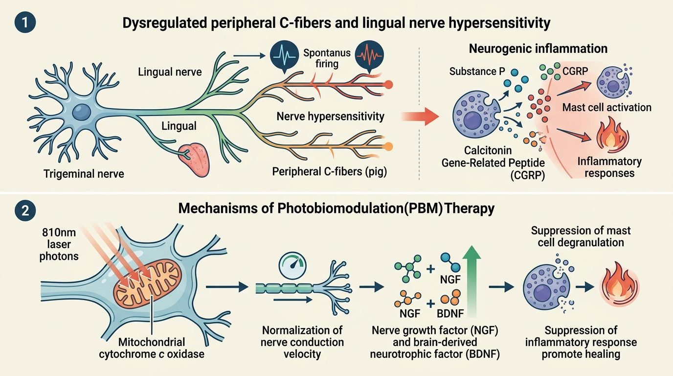
Concurrently, surviving peripheral axons exhibit abnormal ectopic firing, axonal swelling, and dysregulated sensory transduction. Functional magnetic resonance imaging (fMRI) reveals secondary central sensitization, characterized by altered thalamic sensory gating and hypoactivity in the descending endogenous pain-inhibitory dopaminergic pathways within the basal ganglia. At the molecular level, damaged lingual nerve endings overexpress transient receptor potential vanilloid 1 (TRPV1) ion channels and release excessive pain neuro-peptides, notably substance P and calcitonin gene-related peptide (CGRP). These neuro-peptides induce sterile neurogenic inflammation, producing burning sensations akin to scalding liquid exposure in the absence of external physical heat.
Photobiological Mechanisms: Mitochondrial Bioenergetics and Axonal Repair
Photobiomodulation directly targets the cellular bioenergetic crisis underlying peripheral neuropathy. Laser photons in the red (660 nm) and near-infrared (810 nm to 830 nm) spectral bands penetrate the mucosal epithelium, reaching subepithelial lingual nerve plexuses. The primary photoacceptor, mitochondrial cytochrome c oxidase, absorbs these photons, triggering immediate dissociation of inhibitory nitric oxide.
This molecular release accelerates electron transport, elevates mitochondrial membrane potential, and generates a surge in intracellular adenosine triphosphate (ATP) synthesis. Restored ATP availability powers ATP-dependent sodium-potassium pumps (Na+/K+-ATPase) along the axonal lemma. By re-establishing physiological resting membrane potentials, photobiomodulation suppresses spontaneous ectopic action potentials responsible for the burning sensation. Furthermore, low-level laser irradiation upregulates the synthesis of neurotrophic factors, specifically nerve growth factor (NGF) and brain-derived neurotrophic factor (BDNF). These neurotrophins promote Schwann cell migration, stimulate axonal sprouting, and guide structural nerve fiber remyelination across damaged mucosal sensory territories.
Anti-Inflammatory Modulation and Neuro-Peptide Downregulation
Beyond bioenergetic restoration, photobiomodulation dampens the neurochemical drivers of neuropathic hyperalgesia. Photonic stimulation decreases the local transcription of pro-inflammatory cytokines, including tumor necrosis factor-alpha (TNF-a) and interleukin-1 beta (IL-1b), in surrounding mucosal fibroblasts and resident mast cells.
Simultaneously, near-infrared light downregulates the synthesis and exocytosis of substance P and CGRP from sensory nerve terminals. Animal models of peripheral neuropathy confirm that laser irradiation normalizes sensory receptor thresholds, dampening TRPV1 channel hypersensitivity. By extinguishing localized neurogenic inflammation, photonic energy breaks the self-sustaining cycle of sensory sensitization, converting hyperactive peripheral nociceptors into quiescent neural conduits.
Clinical Protocol: Multi-Point Dosimetry and Anatomical Mapping
Achieving predictable clinical remission requires disciplined anatomical mapping and precise optical dosimetry. The clinician must systematically map all anatomical areas affected by dysesthesia, which commonly include the anterior two-thirds of the tongue, the hard palate, and the inner vermilion border of the lower lip.
The standard protocol employs an 810 nm diode laser delivered through a non-defocused biostimulation handpiece with a spot size of 0.8 to 1.0 square centimeter. A multi-point contact technique is executed. The tongue is divided into a geometric grid comprising six to eight discrete points across the dorsal and lateral margins. Each point receives an energy density of 4 to 6 Joules per square centimeter, delivered at a continuous wave power of 100 to 200 milliwatts for twenty to thirty seconds per site. The hard palate receives four to six points of equivalent dosimetry. Treatments are administered twice weekly for four consecutive weeks, transitioning to once weekly for an additional four weeks to solidify neural stabilization. Patients presenting with co-existing dysgeusia (phantom metallic or bitter taste) or perceived xerostomia also experience marked improvements, as near-infrared photons revitalize supporting microvascular networks supplying taste buds in circumvallate and fungiform papillae.
Long-Term Clinical Outcomes: Pain Relief and Quality of Life Metrics
Clinical trials utilizing standardized visual analog scale (VAS) scoring and the Oral Health Impact Profile (OHIP-14) demonstrate profound therapeutic responses following photobiomodulation. Patients treated with active laser therapy achieve mean pain reductions exceeding fifty to seventy percent compared to baseline values, whereas sham-irradiated placebo cohorts show negligible change.
Symptom reduction begins between the third and fifth treatment sessions, with progressive cumulative relief extending across the eight-week treatment arc. Follow-up evaluations at three and six months post-treatment confirm that pain remission remains stable without rebound exacerbation. Furthermore, patients report marked improvements in sleep architecture, mood stabilization, and oral communicative confidence, reflecting the liberation from relentless intraoral pain. Because photobiomodulation operates without systemic absorption, it produces zero pharmacodynamic drug interactions, making it safe for medically complex elderly patients taking polypharmacy regimens. If mild sensory relapse occurs following periods of systemic physical stress or hormonal fluctuations, short booster protocols consisting of three consecutive weekly sessions restore neural latency and maintain patient comfort indefinitely.
Clinical Synthesis for Orofacial Pain Practice
Photobiomodulation therapy shifts the clinical paradigm of burning mouth syndrome from palliative pharmacotherapy to targeted neurobiological regeneration. By restoring axonal ATP synthesis, repolarizing unstable neural membranes, and extinguishing neurogenic cytokine signaling, near-infrared lasers repair the damaged peripheral nerve architecture driving chronic oral burning. Incorporating photobiomodulation empowers orofacial pain specialists and general practitioners to provide reproducible pain relief, restoring psychological comfort and physical wellbeing to patients suffering from this debilitating neuropathic condition.
References
- Arbabi-Kalati, F., et al. (2015). Evaluation of the effect of low-level laser therapy on burning mouth syndrome: a randomized controlled trial. Journal of Clinical and Diagnostic Research, 9(3), ZC54-ZC56.
- Bardellini, E., et al. (2019). Photobiomodulation therapy for the management of burning mouth syndrome: a systematic review and meta-analysis. Oral Diseases, 25(8), 1845-1854.
- De Pedro, M., et al. (2020). Photobiomodulation therapy in burning mouth syndrome: a randomized double-blind clinical trial. Journal of Clinical Medicine, 9(8), 2465.
- Spanemberg, J. C., et al. (2015). Efficacy of low-level laser therapy for the treatment of burning mouth syndrome: a randomized, controlled trial. Lasers in Medical Science, 30(2), 883-889.
- Valenzuela, S., et al. (2017). Burning mouth syndrome: update on pathogenesis, diagnosis, and management. Medicina Oral, Patología Oral y Cirugía Bucal, 22(4), e440-e448.
Leave a Reply