
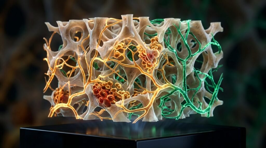
Diabetes mellitus induces microvascular dysfunction, suppresses bone remodeling, and impairs wound healing within the post-extraction alveolar socket. Patients exhibiting poorly controlled type 2 diabetes (glycated hemoglobin HbA1c exceeding 8.0 percent) experience a two- to three-fold higher incidence of post-extraction complications. These complications include dry socket (alveolar osteitis), delayed primary epithelial closure, persistent suppuration, and extensive crestal bone resorption. The underlying pathophysiological cascade involves the chronic accumulation of advanced glycation end-products (AGEs) within the extracellular matrix of alveolar bone and connective tissues. Binding of AGEs to their corresponding multiligand receptor (RAGE) on vascular endothelial cells, macrophages, and osteoblasts triggers persistent oxidative stress, activates nuclear factor-kappa B (NF-kB), and sustains high levels of pro-inflammatory cytokines, specifically tumor necrosis factor-alpha (TNF-a) and interleukin-1 beta (IL-1b). Photobiomodulation (PBM) deploying red and near-infrared optical radiation represents an adjunctive physical therapy designed to stimulate mitochondrial respiration and restore microvascular perfusion. However, PBM cannot magically override the hostile biological milieu of severe, unmanaged hyperglycemia. Safe and reproducible clinical benefits require rigorous dosimetric protocols, structured irradiation geometry, and systemic glycemic stabilization.
The primary cellular mechanism by which PBM counteracts diabetic wound stalling operates through mitochondrial chromophore excitation. Photons in the optical window of 630 nm to 850 nm are preferentially absorbed by cytochrome c oxidase (Unit IV of the mitochondrial electron transport chain). In diabetic tissues, persistent oxidative stress disrupts electron transfer, leading to uncoupled oxidative phosphorylation and depleted adenosine triphosphate (ATP) reserves. Photon absorption prompts photodissociation of inhibitory nitric oxide from the binuclear heme centers of cytochrome c oxidase. This dissociation restores the catalytic activity of the enzyme, elevates mitochondrial membrane potential, and drives energetic ATP synthesis in hypoperfused alveolar fibroblasts and osteoprogenitor cells. Concurrently, transient, controlled low-level bursts of reactive oxygen species (ROS) activate downstream transcription factors, including hypoxia-inducible factor-1 alpha (HIF-1a), which orchestrates vascular endothelial growth factor (VEGF) synthesis and initiates robust capillary sprouting across the hypovascular extraction clot.
Alveolar bone repair requires coordinated coupling between osteoclasts and osteoblasts, a balance pathologically disrupted in diabetic microenvironments. Hyperglycemia skews the ratio of receptor activator of nuclear factor-kappa B ligand (RANKL) to osteoprotegerin (OPG), favoring uncontrolled osteoclastogenesis and persistent osteoclastic bone resorption along the alveolar margins. Furthermore, osteoblast apoptosis is markedly accelerated by AGE-RAGE signaling. Laboratory animal models and human bone biopsy investigations indicate that near-infrared PBM (specifically 808 nm to 830 nm) down-regulates osteoclastogenic RANKL secretion from osteocytes while up-regulating OPG expression in alveolar osteoblasts. This biochemical normalization reduces peripheral crestal bone loss during the initial thirty days following extraction. Histomorphometric analyses demonstrate accelerated trabecular bone deposition, increased mineralized bone volume fraction, and denser lamellar organization within the apical and middle thirds of laser-treated diabetic extraction sockets.
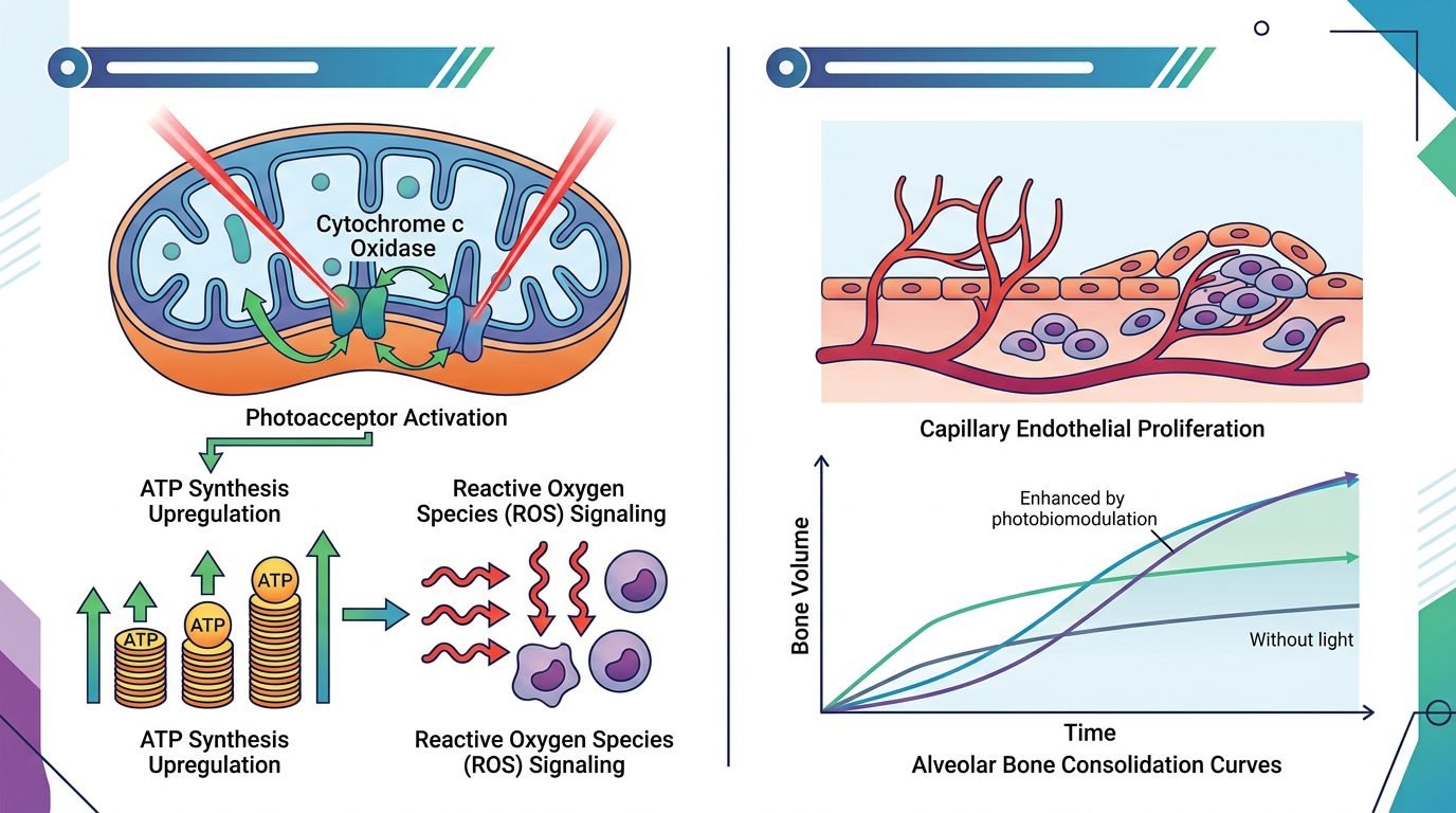
Dosimetric calibration determines the boundary between therapeutic biostimulation and photothermal tissue inhibition. In accordance with the Arndt-Schulz biphasic dose-response curve, insufficient energy densities fail to overcome diabetic bioenergetic deficits, whereas excessive fluences produce cellular bioinhibition, intracellular hyperthermia, and oxidative cytotoxicity. For post-extraction alveolar sockets in diabetic patients, clinical protocols require wavelengths capable of adequate optical penetration through thick cortical plates and organized blood clots. Red wavelengths (660 nm) exhibit high absorption in superficial hemoglobin and melanin, making them ideal for accelerating superficial epithelial seal closure across the socket orifice. Conversely, near-infrared wavelengths (808 nm to 830 nm) display lower scattering and minimal water absorption, delivering therapeutic photon densities down into deep medullary trabeculae.
The optimal clinical delivery protocol combines intra-socket and trans-mucosal irradiation patterns. Immediately following atraumatic extraction, thorough curettage of chronic granulation tissue, and copiously rinsing with sterile saline, the laser probe is introduced without contacting the fragile blood clot. For superficial mucosal biostimulation, a 660 nm diode laser is applied at an energy density of four to six joules per square centimeter with an output power of fifty milliwatts over the mucosal margins. To stimulate deep alveolar osteogenesis, an 810 nm near-infrared diode laser is applied to the buccal and lingual cortical plates at an energy density of six to eight joules per square centimeter per point, utilizing a spot size of approximately 0.5 to 1.0 square centimeter and an irradiance between one hundred and two hundred milliwatts per square centimeter. Treatment sessions must follow a structured temporal schedule: immediately post-extraction (Day 0), followed by recall applications on Day 2, Day 4, and Day 7.
Longitudinal clinical outcome assessments provide realistic boundaries for expected treatment success. In a randomized controlled clinical trial comparing PBM to sham irradiation in poorly controlled type 2 diabetic patients undergoing premolar extractions, complete epithelial soft tissue closure occurred significantly earlier in the laser cohort (mean 11.2 days versus 18.7 days). Radiographic pixel density analysis at eight and twelve weeks revealed higher bone mineral fill in the coronal third of the extraction sockets. However, systemic glycemic status fundamentally controls the magnitude of the clinical response. In patients with severe, unmanaged hyperglycemia characterized by HbA1c levels exceeding 10.0 percent, the clinical benefits of PBM were substantially attenuated. Massive systemic microvascular damage and pervasive circulating glycosylated proteins blunted local cellular responsiveness. Clinicians must educate patients that PBM represents an adjunctive physical booster, not an alternative to medical glycemic stabilization, dietary adherence, and oral antimicrobial maintenance.
Comparative clinical analyses examine whether PBM can substitute for prophylactic local antibiotic therapy in compromised diabetic sockets. Placing minocycline or tetracycline ointments directly into fresh extraction sockets presents documented risks of foreign-body granulomatous reactions, cytotoxic inhibition of migrating gingival fibroblasts, and the emergence of resistant oral strains. Photobiomodulation provides non-pharmacological antimicrobial suppression indirectly: by promoting rapid microvascular reperfusion and elevating endogenous neutrophil oxidative burst activity within the socket lumen, optical irradiation limits bacterial colonisation without chemical side effects. When paired with post-operative chlorhexidine rinses (0.12 percent alcohol-free) twice daily for seven days, PBM achieves comparable or superior socket infection prevention relative to topical antibiotic pastes, while preserving local cellular osteogenic viability.
Surgical technique remains the baseline determinant of post-extraction healing. PBM cannot compensate for traumatic bone expansion, fractured cortical plates, or neglected bacterial residues. Extractions in diabetic patients must be performed with meticulous periotomes, cross-cut fissure burs with abundant saline irrigation, and minimal mucoperiosteal reflection to protect existing periosteal microvascular networks. The placement of resorbable collagen sponges or platelet-rich fibrin (PRF) scaffolds inside the socket stabilizes the initial fibrin clot, providing an ideal cellular matrix for photonic stimulation.
Regarding clinical implementation, photobiomodulation utilizing red and near-infrared wavelengths provides a biologically grounded, non-invasive therapeutic intervention to accelerate soft tissue epithelialization and stimulate bone fill in diabetic post-extraction sockets. The clinical protocol demands careful energy titration, strict adherence to non-thermal fluences, and structured application across the initial post-operative week. When combined with minimally invasive exodontia, socket stabilization, and physician-coordinated glycemic control, PBM effectively lowers the risk of post-surgical alveolar complications and preserves critical alveolar ridge architecture.
Leave a Reply