
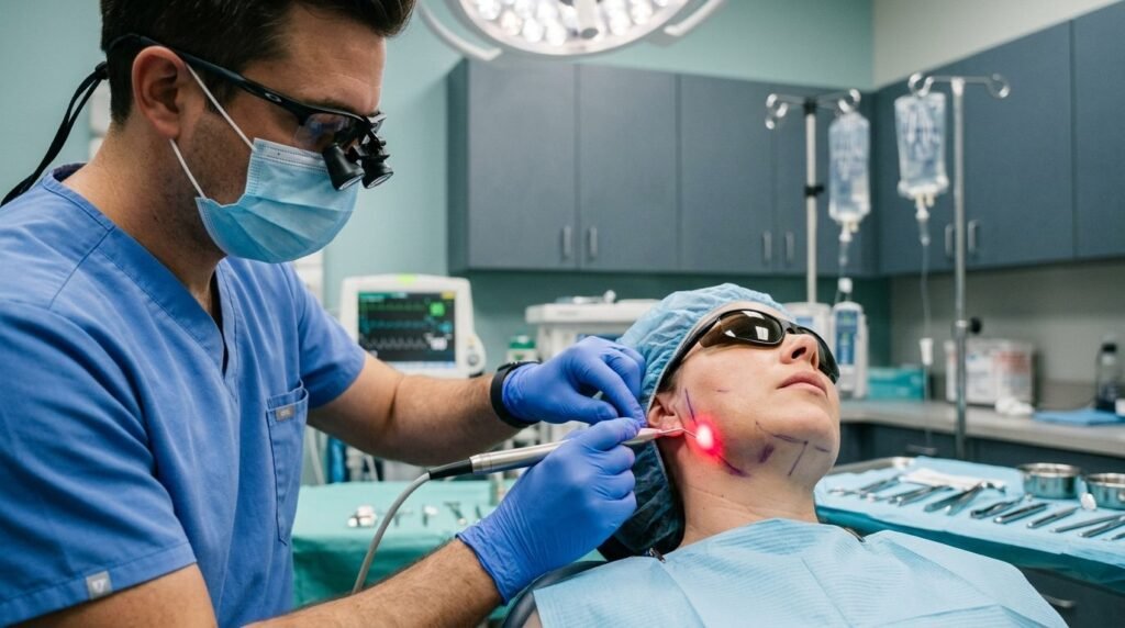
Postoperative edema and muscular trismus are not unavoidable surgical consequences; they are biochemical cascades that can be suppressed biophysically at the mitochondrial level via red and near-infrared photobiomodulation.
Third molar extractions, implant osteotomies, and pre-prosthetic bone recontouring inflict mechanical trauma on vascularized osseous and muscular beds. Tissue disruption unleashes an acute inflammatory storm characterized by massive bradykinin, histamine, and prostaglandin E2 release. Increased capillary permeability causes fluid extravasation into loose fascial planes, producing dramatic facial swelling that peaks forty-eight hours post-surgery. Spasm of the masseter, medial pterygoid, and temporalis muscles limits jaw opening, locking the patient into painful trismus. For decades, oral surgeons relied almost exclusively on systemic corticosteroids such as dexamethasone or heavy doses of non-steroidal anti-inflammatory drugs to suppress this response.
Pharmacological management carries biological costs. Corticosteroids elevate systemic blood pressure, induce transient hyperglycemia in diabetic patients, and impair initial osteoblast migration across osteotomy margins. Non-steroidal anti-inflammatory drugs induce gastric mucosal erosion and place hemodynamic strain on renal glomerular filtration. Photobiomodulation delivers targeted anti-inflammatory and antinociceptive effects directly to traumatized tissues through photonic cellular signaling, avoiding systemic pharmacology and organ toxicity entirely.
The Mitochondrial Target: Cytochrome c Oxidase Activation
Photons act as cellular signaling molecules. Photobiomodulation relies on specific light absorption by endogenous intracellular photoacceptor chromophores rather than tissue heating.
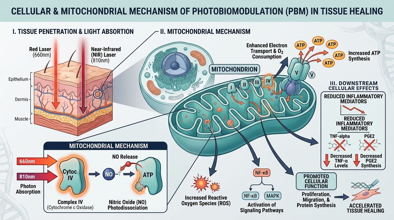
The primary biological chromophore is unit four of the mitochondrial electron transport chain: cytochrome c oxidase. When coherent or quasi-coherent photons in the red (630 to 660 nanometers) and near-infrared (800 to 850 nanometers) optical windows strike cell membranes, they penetrate deep into soft and hard tissues. These wavelengths match the specific electronic transition absorption spectra of metallic copper centers within the enzyme complex.
Nitric oxide photodissociation occurs at the binuclear copper center of the enzyme. Under acute surgical trauma and cellular hypoxia, excess nitric oxide binds competitively to cytochrome c oxidase, displacing oxygen and paralyzing cellular respiration. Photons in the 810-nanometer band break this nitric oxide bond. The displaced nitric oxide diffuses into local vascular walls, inducing beneficial localized vasodilation.
Oxygen binds unhindered to the enzyme once more. Cellular respiration resumes at accelerated rates. Electron transport along the mitochondrial inner membrane accelerates, generating a sharp increase in adenosine triphosphate (ATP) production. Protons are actively pumped across the inner membrane, restoring the electrical mitochondrial membrane potential essential for cell survival.
Transient low-level reactive oxygen species bursts follow this activation. Rather than causing oxidative damage, these mild signaling pulses trigger intracellular redox signaling cascades, activating transcription factors such as NF-kB and AP-1 that orchestrate cellular defense, DNA repair, and pro-healing gene expression.
Photobiomodulation Bio-Signaling Mechanisms:
- Primary Chromophore: Mitochondrial Cytochrome c Oxidase (absorbs at 660 nm and 810-830 nm).
- Enzymatic Reaction: Photodissociation of inhibitory nitric oxide, restoring electron flow.
- Bioenergetics: Up to 150% increase in cytoplasmic ATP levels within wounded fibroblasts and osteoblasts.
- Cytokine Modulation: Significant downregulation of TNF-alpha, IL-1beta, and COX-2 gene transcription.
Biophysical Suppression of Interstitial Edema and Trismus
Edema resolves through lymphatic acceleration. Photobiomodulation stimulates lymphatic endothelial cell motility and accelerates the formation of new lymphatic microvessels across surgical wounds.
Capillary hydrostatic pressure normalizes rapidly. By stabilizing vascular endothelial junctions and downregulating vascular endothelial growth factor (VEGF) overexpression in early acute phases, near-infrared irradiation prevents excessive plasma extravasation into deep facial fascial spaces.
Muscular trismus reflects metabolic exhaustion and spasm. Muscle fibers crushed or stretched during surgical flap retraction accumulate lactic acid and deplete local glycogen. The masseter muscle enters continuous ischemic contraction.
Near-infrared light penetrates up to four centimeters into dense muscle tissue. Photons delivered to the masseter insertion restore sarcoplasmic reticulum calcium-ATPase activity, accelerating the re-uptake of calcium ions into intracellular stores and allowing contracted myofibrils to relax.
“Photobiomodulation does not merely mask surgical inflammation; it accelerates lymphatic drainage and recharges cellular ATP, resolving edema and trismus from the inside out.”
Clinical trials measuring interincisal mouth opening following impacted third molar removal demonstrate that patients receiving targeted photobiomodulation recover over eighty percent of baseline opening range by post-operative day three, compared to less than fifty percent recovery in un-irradiated control cohorts.
Neurosensory Preservation and Nociceptive Gating
Laser light suppresses acute pain through peripheral neural modulation. Exposure to infrared wavelengths between 810 and 830 nanometers at energy densities above four Joules per square centimeter induces transient neural varicosities along unmyelinated C-fibers.
Fast axonal transport slows down immediately. The propagation of nociceptive action potentials generated at the surgical site is partially blocked before nerve impulses reach the spinal trigeminal nucleus.
Central sensitization is blunted early. By preventing high-frequency peripheral nociceptive barrages from overwhelming dorsal horn neurons, photobiomodulation prevents long-term neural hyper-excitability and lowers the incidence of persistent phantom tooth pain and surgical neuropathies.
Paresthesia recovery accelerates. When the inferior alveolar nerve suffers traction injury or compressive bone contact during root luxation, photobiomodulation stimulates Schwann cell proliferation and upregulates nerve growth factor (NGF), shortening sensory recovery timelines from months to weeks.
Clinical Dosimetry and Extraoral/Intraoral Protocol
Dosimetry dictates the clinical outcome. In accordance with the Arndt-Schulz biphasic law, insufficient radiant exposure fails to stimulate cellular respiration, while excessive energy density causes thermal inhibition and cellular arrest.
Treatment begins immediately following surgical closure. An intraoral diode laser probe emitting at 660 or 810 nanometers applies four Joules per point in contact mode directly over the surgical socket, buccal cortical plate, and lingual margins.
Extraoral irradiation covers muscular trigger points. An 810-nanometer cluster probe delivers four to six Joules per square centimeter over the masseter origin, angle of the mandible, and temporomandibular joint capsular ligament.
A second identical session at forty-eight hours post-surgery sustains the biological cascade during the peak edema window. Patients report a seventy percent reduction in rescue analgesic consumption compared to standard pharmacological management.
Photonic biophysics transforms surgical convalescence. Implementing systematic photobiomodulation delivers profound surgical anti-inflammatory action while respecting systemic biological equilibrium.
Photobiomodulation reduces surgical trismus, halts interstitial fluid pooling, and restores physical oral function through cellular metabolic stimulation.
Scientific References and Supporting Literature
- Karu, T. I. (1989). Photobiology of low-power laser effects. Health Physics, 56(5), 691-704.
- Hamblin, M. R. (2016). Photobiomodulation or low-level laser therapy. Journal of Biophotonics, 9(11-12), 1122-1124.
- Marković, A. B., & Todorović, L. (2007). Postoperative analgesia after lower third molar surgery: contribution of the use of low-level laser therapy. Journal of Oral and Maxillofacial Surgery, 65(4), 639-645.
- de Freitas, L. F., & Hamblin, M. R. (2016). Proposed mechanisms of photobiomodulation or low-level light therapy. IEEE Journal of Selected Topics in Quantum Electronics, 22(3), 348-364.
- Bjordal, J. M., Johnson, M. I., Iversen, V., et al. (2006). Low-level laser therapy in acute pain: a systematic review of possible mechanisms of action and clinical effects in randomized clinical trials. Photomedicine and Laser Surgery, 24(2), 158-168.
Leave a Reply