
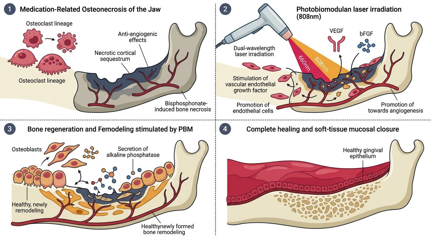
Photobiomodulation therapy combined with surgical laser debridement halts necrotic progression and achieves complete mucosal closure in medication-related osteonecrosis of the jaw where conventional medical therapies fail. Medication-related osteonecrosis of the jaw (MRONJ) occurs primarily in oncology and osteoporosis patients receiving high-potency antiresorptive agents, such as zoledronic acid and denosumab, or antiangiogenic therapies. These medications suppress osteoclastic bone remodeling, inhibit endothelial cell proliferation, and cause microvascular thrombosis within cortical jawbones. Conservative antimicrobial rinses and antibiotics achieve mucosal closure in under twenty percent of advanced lesions, whereas radical surgery risks fractures and severe deformity. Near-infrared photobiomodulation stimulates mitochondrial ATP synthesis, promotes neoangiogenesis, and upregulates osteoblastic differentiation, transforming avascular necrotic bone into an active regenerative environment.
Pathophysiology of MRONJ: The Suppression of Bone Remodeling and Angiogenesis
Bisphosphonates incorporate directly into calcium hydroxyapatite crystals within alveolar bone, where they persist for decades. When osteoclasts ingest bisphosphonate-laden mineral during bone turnover, the nitrogen-containing molecule inhibits farnesyl pyrophosphate synthase in the mevalonate pathway. This disruption prevents protein prenylation, causing loss of the osteoclast ruffled border and triggering rapid apoptotic cellular death.
Denosumab, a human monoclonal antibody targeting receptor activator of nuclear factor-kappa B ligand (RANKL), achieves similar osteoclast suppression by blocking osteoclastogenesis and differentiation. Concurrently, antiresorptive agents exert direct antiangiogenic toxicity on oral mucosal tissues, down-regulating vascular endothelial growth factor (VEGF) receptors and inhibiting capillary tubule formation. The human jawbones, possessing high physiological bone turnover rates due to continuous masticatory stresses, cannot repair micro-damage. Tooth extractions or denture pressure ulcers expose hypovascular, non-vital bone to oral microbial biofilms. Microbial colonization by Actinomyces and oral streptococci leads to persistent osteomyelitis, bone necrosis, and painful soft-tissue breakdown.
Photobiological Mechanisms: Angiogenesis and Mitochondrial Activation
The core therapeutic challenge in MRONJ is overcoming severe local tissue ischemia. Photobiomodulation delivers red (660 nm) and near-infrared (810 nm) photons that target cytochrome c oxidase within damaged endothelial cells, fibroblasts, and osteoblasts. Photon absorption stimulates the mitochondrial respiratory chain, increasing adenosine triphosphate production and restoring cellular membrane potential.
Irradiation triggers controlled reactive oxygen signaling that activates hypoxia-inducible factor 1-alpha, driving VEGF and fibroblast growth factor secretion for neoangiogenesis. In vitro capillary tube formation assays confirm that near-infrared laser irradiation stimulates migration of endothelial progenitor cells across ischemic tissue margins, fostering robust capillary sprouting. Concurrently, photobiomodulation stimulates alkaline phosphatase activity and osteocalcin expression in local mesenchymal stem cells, promoting bone remodeling along viable osteotomy borders.
Clinical Protocol: Staging, Laser Selection, and Conservative Pre-Conditioning
Clinical management must follow the staging system established by the American Association of Oral and Maxillofacial Surgeons (AAOMS). Stage Zero presents with non-specific bone pain and radiographic sclerosis without exposed bone; Stage One features exposed necrotic bone without pain or infection; Stage Two exhibits exposed bone with pain and purulent erythema; Stage Three presents with extensive exposed bone extending beyond the alveolar ridge, extraoral fistulae, or pathological fractures.
Treatment begins with a conservative pre-conditioning phase designed to reduce local bacterial load and stimulate baseline vascularity. Chlorhexidine 0.12 percent rinses are combined with systemic amoxicillin with clavulanate (or clindamycin in penicillin-allergic patients). Photobiomodulation is initiated chairside using an 810 nm diode laser in continuous wave mode. Using a non-contact handpiece, the laser delivers 4 to 6 Joules per square centimeter across the necrotic bone bed and adjacent erythematous mucosa. Sessions occur twice weekly for three to four weeks. In Stage One lesions, this non-surgical protocol frequently stimulates peripheral mucosal migration, resulting in complete spontaneous soft-tissue coverage over non-vital bone.
Surgical Laser Debridement: Erbium Laser Resection and Fibrin Sealing
For refractory Stage Two and Stage Three lesions where non-surgical protocols fail to achieve closure, surgical debridement using Er:YAG (2,940 nm) or Er,Cr:YSGG (2,780 nm) lasers represents the gold standard. Conventional rotary bur debridement generates frictional heat and deposits a thick smear layer, creating additional microvascular thrombosis that extends bone necrosis.
The erbium laser operates via thermomechanical micro-explosions under continuous sterile water spray, abating necrotic bone without thermal coagulation. The clinician resects non-vital bone until bleeding, viable trabecular bleeding points appear. Er:YAG lasers decontaminate bone margins by vaporizing microbial biofilms sequestered within Haversian canals. Immediately following osseous debridement, low-level 810 nm laser irradiation is applied directly to the bleeding bone bed at 4 Joules per square centimeter to stimulate local microcirculation. Autologous platelet-rich fibrin (PRF) membranes are placed over the defect, and mucosal flaps are mobilized and closed tension-free using monofilament sutures.
Postoperative Photobiomodulation and Maintenance Regimens
Photobiomodulation must continue postoperatively to support flap viability and accelerate mucosal re-epithelialization. Transcutaneous and intraoral irradiation is administered at 4 Joules per square centimeter on postoperative days one, three, seven, fourteen, and twenty-one.
Monitoring tissue healing requires clinical and radiographic documentation. Near-infrared photobiomodulation relieves acute postoperative pain, decreasing analgesic requirements. By stimulating fibroblast collagen synthesis and reducing local inflammatory edema, photonic therapy prevents mucosal suture dehiscence. Clinical trials demonstrate that combining surgical erbium debridement with adjuvant photobiomodulation achieves complete mucosal healing rates exceeding eighty-five percent, compared to less than fifty percent achieved with conventional surgical techniques.
Interprofessional Coordination with Medical Oncology
Managing patients with MRONJ requires close interprofessional communication with treating medical oncologists and endocrinologists. Dental clinicians must never unilaterally discontinue oncological antiresorptive or antiangiogenic therapy. Drug holidays for oral bisphosphonates in osteoporosis may be considered in consultation with physicians, but temporary cessation of intravenous antiresorptive therapy in cancer patients with skeletal metastases provides negligible short-term oral benefit while risking catastrophic skeletal-related events, including spinal cord compression and pathological fractures.
Pre-treatment dental screening before initiating antiresorptive therapy represents the most effective preventive strategy. All non-restorable teeth, advanced periodontal lesions, and active oral infections must be surgically resolved, allowing complete mucosal healing before oncological infusions begin. When osteonecrosis does develop, photobiomodulation provides a predictable, non-invasive therapeutic modality that restores oral health while allowing essential oncological therapies to proceed safely.
Clinical Synthesis for Oral and Maxillofacial Practice
Photobiomodulation therapy shifts MRONJ management from therapeutic nihilism to predictable tissue regeneration. By restoring cellular bioenergetics, accelerating neoangiogenesis, and enabling atraumatic surgical debridement, bio-photonic modalities resolve necrotic lesions that resist conventional medicine. Mastery of optical dosimetry and combined wavelength protocols empowers the dental team to achieve complete mucosal closure, alleviate debilitating pain, and protect the systemic wellbeing of medically complex patients.
References
- Ruggiero, S. L., et al. (2022). Medication-related osteonecrosis of the jaw: 2022 update. Journal of Oral and Maxillofacial Surgery, 80(5), 920-943.
- Vescovi, P., et al. (2012). Nd:YAG laser biostimulation in the treatment of bisphosphonate-associated osteonecrosis of the jaw: clinical experience in 28 cases. Photomedicine and Laser Surgery, 26(1), 37-46.
- Atasoy, K. T., et al. (2017). Photobiomodulation therapy in the treatment of medication-related osteonecrosis of the jaw: a systematic review. Photomedicine and Laser Surgery, 35(12), 654-663.
- Scoletta, M., et al. (2010). Low-level laser therapy in the management of bisphosphonate-induced osteonecrosis of the jaws: a retrospective study of 20 patients. Lasers in Medical Science, 25(3), 441-447.
- Stübinger, S., et al. (2009). The use of Er:YAG laser in the treatment of bisphosphonate-related osteonecrosis of the jaws. Lasers in Surgery and Medicine, 41(5), 350-357.
Leave a Reply