
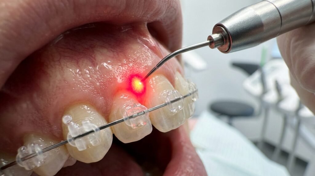
Mechanical orthodontic force application induces localized aseptic inflammation within the periodontal ligament, initiating alveolar bone remodeling alongside significant physical discomfort. When mechanical archwires or clear aligners engage teeth, compressive stresses on the periodontal ligament restrict microvascular capillary perfusion, creating focal ischemic necrosis and hyalinization. Concurrently, sensory nerve terminals release substance P, bradykinin, and prostaglandins, provoking intense ischemic pain during mastication that peaks twenty-four to forty-eight hours post-activation. Systemic non-steroidal anti-inflammatory analgesics taken to manage this discomfort frequently inhibit cyclooxygenase enzymes, inadvertently blunting osteoclastic bone resorption and slowing orthodontic velocity. Photobiomodulation therapy deployed at red (660 nm) and near-infrared (810 to 830 nm) wavelengths presents a non-pharmacological biostimulative modality that simultaneously accelerates bone turnover and relieves acute periodontal pain without arresting tooth movement.
Cellular and Molecular Dynamics of Orthodontic Biomechanics
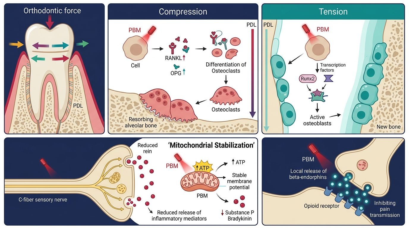
Orthodontic tooth movement depends fundamentally on coordinated coupled bone remodeling governed by the compression-tension paradigm. On the compression side of the periodontal ligament, mechanical force triggers localized hypoxia and cellular stress. Periodontal ligament fibroblasts and osteocytes upregulate receptor activator of nuclear factor-kappa B ligand (RANKL) while downregulating osteoprotegerin (OPG). The elevated RANKL/OPG ratio binds to RANK receptors on circulating monocytic precursors, stimulating their differentiation into multinucleated osteoclasts that resorb the compressive alveolar bone wall.
Conversely, on the tension side, mechanical stretching stimulates periodontal fibroblasts and mesenchymal stem cells to upregulate runt-related transcription factor 2 (Runx2), alkaline phosphatase, and osteocalcin. These osteogenic signaling cascades drive osteoblastic matrix deposition and subsequent mineralization, anchoring the tooth in its newly established spatial position. However, if hyalinized necrotic zones on the compression side are excessively large, tooth displacement pauses until adjacent scavenger macrophages clear cellular debris through undermining resorption, prolonging overall clinical treatment timelines.
Mitochondrial Photobiomodulation and Accelerated Bone Remodeling
Photobiomodulation delivers coherent photons that penetrate cortical and trabecular alveolar bone plates, reaching target cells within the stressed periodontal ligament space. At the cellular level, near-infrared photons (810 to 830 nm) are absorbed by cytochrome c oxidase within unit four of the mitochondrial respiratory chain. This photo-activation accelerates electron transfer rates, elevating mitochondrial membrane potentials and dramatically increasing adenosine triphosphate production. Intracellular cyclic adenosine monophosphate and reactive oxygen species surge within physiological thresholds, triggering nuclear factor-kappa B and activator protein 1 transcription factors.
This bioenergetic surge accelerates biological turnover on both sides of the moving tooth. On the compression side, laser-irradiated periodontal fibroblasts exhibit heightened and expedited secretion of RANKL, promoting faster osteoclastogenesis and accelerating the enzymatic breakdown of hyalinized necrotic tissue. On the tension side, photobiostimulated osteoblasts demonstrate enhanced proliferation, increased alkaline phosphatase activity, and rapid collagenous osteoid synthesis. Multiple randomized split-mouth clinical trials demonstrate that calibrated photobiomodulation accelerates canine retraction and overall alignment phases by 30 to 40 percent compared to conventional mechanical mechanics alone.
Electrophysiological and Biochemical Mechanisms of Analgesia
The simultaneous analgesic action of photobiomodulation addresses the primary complaint of orthodontic patients: post-activation pain. Mechanical pressure on the periodontal ligament compresses sensory nerve bundles, triggering local acidosis, neurogenic inflammation, and unmyelinated C-fiber hyperexcitability. Traditional non-steroidal anti-inflammatory drugs inhibit prostaglandin synthesis systemically, which relieves discomfort but simultaneously impairs the inflammatory cascade required for osteoclast recruitment, thereby retarding tooth movement.
Photobiomodulation achieves profound localized analgesia through distinct, non-destructive neurochemical pathways that leave bone remodeling cascades entirely unimpaired. Low-level near-infrared irradiation stabilizes mitochondrial membrane potentials in primary sensory neurons, inducing reversible micro-tubular conduction blocks that suppress fast nociceptive action potential propagation. Concurrently, laser energy suppresses localized concentrations of bradykinin, histamine, and substance P within the compressed periodontal ligament. Improved microcirculatory blood flow washes away acidic metabolic waste products, restoring normal extracellular pH. Additionally, photonic stimulation prompts localized release of beta-endorphins, significantly lowering visual analog pain scores during the first three days following appliance adjustment.
Clinical Dosimetry and Operatory Application Protocols
Achieving predictable clinical results requires rigorous attention to optical energy density, power output, and anatomical delivery geometry. Near-infrared wavelengths between 808 and 830 nanometers are preferred for orthodontic acceleration due to their superior photon penetration depth through thick cortical plates. In contrast, 660 nm red light is highly effective for superficial analgesia and mucosal healing around micro-implants and auxiliary attachments.
Therapeutic energy density should range between 4 and 8 Joules per square centimeter per point, delivered in continuous-wave mode with power settings between 100 and 200 milliwatts. The laser probe must be positioned perpendicular to the alveolar mucosa overlying the root of the targeted tooth, maintaining light contact without blanching the tissue. For single-rooted teeth, irradiation should cover four points: cervical buccal, apical buccal, cervical lingual or palatal, and apical lingual. For multi-rooted molars, irradiation points expand to six to eight locations to ensure uniform energy distribution across the root surface area. Treatments administered immediately following archwire ligation, repeated at twenty-four hours and weekly thereafter, maintain accelerated cellular metabolism.
At-Home Extraoral versus In-Office Intraoral Systems
The practical integration of photobiomodulation into orthodontic practice encompasses both high-power in-office intraoral laser devices and low-power at-home extraoral LED arrays. In-office delivery utilizes focused fiber-optic handpieces operated by the clinician or trained auxiliary immediately following force application. This approach ensures strict quality control over dosimetric delivery, correct anatomical angulation, and verified energy density. However, because photobiomodulation effects peak biologically within twenty-four to forty-eight hours, office-only visits require multiple weekly appointments that can challenge patient schedules.
Conversely, customized extraoral LED devices emitting 850 nm light allow patients to self-administer daily five to ten-minute treatments at home. While LED arrays exhibit higher beam divergence and lower photon penetration compared to coherent lasers, daily home compliance compensates for lower peak energy densities. Regardless of delivery hardware, practitioner guidance, standardized dosimetry, and regular monitoring remain indispensable for maximizing clinical velocity and safety.
Significance for Modern Humanized Orthodontics
Deploying photobiomodulation redefines orthodontic care by aligning mechanical physics with host biological responses. Accelerating tooth movement while eradicating treatment-induced pain transforms the patient experience, substantially reducing treatment dropout rates and root resorption risks associated with prolonged appliance wear. By harnessing targeted photobiology, orthodontists achieve rapid, comfortable, and biologically sound tooth alignment.
References
- Gkantidis, N., et al. (2014). Effectiveness of methods used to accelerate orthodontic tooth movement: a systematic review. European Journal of Orthodontics, 36(5), 552-568.
- Bartzela, T., et al. (2009). Cellular and molecular mechanisms in orthodontic tooth movement: an update. American Journal of Orthodontics and Dentofacial Orthopedics, 135(5), 616-624.
- Marques, L. S., et al. (2015). Low-level laser therapy accelerates orthodontic tooth movement: a meta-analysis. Lasers in Medical Science, 30(2), 795-802.
- Qamruddin, I., et al. (2017). The effect of photobiomodulation on the rate of orthodontic tooth movement and pain: a systematic review and meta-analysis. Journal of Orthodontics, 44(4), 282-293.
- Sousa, M. V., et al. (2014). Effect of low-level laser therapy on orthodontic tooth movement: a randomized clinical trial. Lasers in Medical Science, 29(6), 1913-1919.
Leave a Reply