
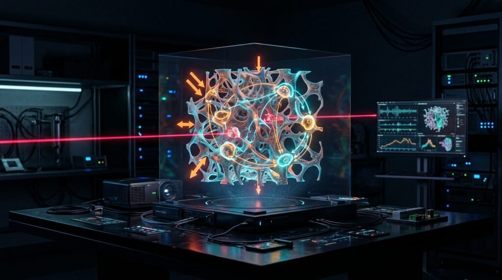
Photobiomodulation therapy accelerates orthodontic tooth movement by stimulating ATP production and modulating osteoclastogenic cytokine cascades within the periodontal ligament without inducing root resorption. Traditional orthodontic mechanics depend on mechanical forces to induce sterile inflammation, generating localized hypoxia, capillary compression, and fluid shear stress across alveolar bone. Biological bone turnover represents the rate-limiting step in comprehensive orthodontic therapy: osteoclastic bone resorption on the compression side must precede osteoblastic bone formation on the tension side. When biological remodeling lags behind mechanical force application, patients experience prolonged treatment durations, elevated risks of external apical root resorption, and severe post-activation ischemic pain. Applying near-infrared photobiomodulation (810 nm to 830 nm) delivers optical energy directly to alveolar osteocytes and periodontal fibroblasts. This photonic stimulus upregulates the RANKL/OPG signaling pathway, accelerating tooth transit velocities by thirty to forty percent while relieving acute post-adjustment pain.
Cellular Bioenergetics and Mitochondrial Photon Absorption
Photobiomodulation operates via non-thermal photophysical mechanisms in target tissues. Near-infrared photons penetrate oral mucosal soft tissues and cortical alveolar bone plates to reach the periodontal ligament space. Within this mechanical-vascular interface, the primary photoacceptor is cytochrome c oxidase, Unit Four of the mitochondrial respiratory electron transport chain.
Photon absorption displaces inhibitory nitric oxide from the binuclear catalytic centers of cytochrome c oxidase. This displacement restores normal electron transport, increases mitochondrial membrane potential, and amplifies adenosine triphosphate (ATP) synthesis within stressed periodontal ligament fibroblasts. Concurrently, transient, low-amplitude reactive oxygen species bursts activate redox-sensitive transcription factors, notably nuclear factor-kappa B (NF-kB) and hypoxia-inducible factor 1-alpha (HIF-1a). These transcription factors initiate cellular proliferation, accelerate local capillary neoangiogenesis, and release growth factors that prime alveolar bone margins for rapid cellular remodeling.
Molecular Modulation: RANKL/OPG Dynamics Across Tension and Compression Zones
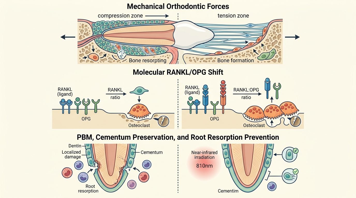
Orthodontic translation requires precise spatial orchestration between bone breakdown and bone apposition. On the compression side of a moving tooth, sustained mechanical pressure occludes microcapillaries, producing localized tissue hyalinization. Before tooth movement can occur, macrophage scavenger cells must clear necrotic hyalinized tissue, and osteoclasts must differentiate to resorb the lamina dura.
Photobiomodulation fundamentally alters this biological timeline. In vitro and animal studies demonstrate that 810 nm laser irradiation stimulates periodontal ligament cells on the compression side to upregulate expression of receptor activator of nuclear factor-kappa B ligand (RANKL). Elevated RANKL binds to RANK receptors on circulating monocyte-macrophage precursors, accelerating osteoclastogenesis and eliminating the hyalinized zone days earlier than conventional mechanics allow. Conversely, on the tension side where bone formation is required, photobiomodulation downregulates RANKL while upregulating osteoprotegerin (OPG), a decoy receptor that prevents excessive bone resorption. Simultaneously, photonic stimulation enhances alkaline phosphatase activity and osteocalcin expression in local osteoblasts, accelerating mature trabecular bone deposition behind the advancing root.
Treatment Acceleration: Clinical Evidence in Canine Retraction and Alignment
Accelerating orthodontic tooth movement reduces overall treatment time, diminishing the window during which patients remain vulnerable to enamel demineralization, white spot lesions, and caries. Randomized controlled trials evaluating canine retraction following premolar extractions provide reliable clinical models for measuring tooth movement velocity.
In split-mouth clinical trials, quadrants receiving adjuvant photobiomodulation exhibit canine retraction rates of 1.2 to 1.6 millimeters per month, compared to 0.8 to 1.0 millimeter per month in control quadrants. This represents a thirty to fifty percent acceleration in canine movement velocity. Similar kinetic gains occur during the initial leveling and alignment phases, where Little’s Irregularity Index resolves significantly faster in laser-treated arches. Importantly, micro-computed tomography and cone-beam volumetric imaging confirm that photobiomodulated teeth show no increase in external apical root resorption compared to non-irradiated controls. The accelerated transit time shortens the duration of active force application, protecting root apex integrity.
Analgesic Mechanisms: Alleviating Post-Adjustment Pain
Acute pain following orthodontic appliance placement or archwire activation is a leading cause of patient dissatisfaction and treatment discontinuation. Pain peaks between 24 and 48 hours post-adjustment, driven by microvascular ischemia, local acidosis, and excessive release of pro-inflammatory mediators, including substance P, prostaglandins, and bradykinin in the periodontal ligament.
Applying photobiomodulation immediately following archwire engagement provides potent chairside analgesia. Low-level near-infrared irradiation blocks the transmission of pain signals along unmyelinated C-fibers by decreasing mitochondrial membrane potential in sensory neurons, transiently suppressing peripheral action potentials. Concurrently, photonic energy reduces the local synthesis of prostaglandin E2 (PGE2) by downregulating cyclooxygenase-2 (COX-2) enzyme expression. Clinical trials show that patients receiving laser therapy report significantly lower visual analog scale pain scores at 6, 24, and 48 hours compared to sham-irradiated controls, eliminating the need for systemic non-steroidal anti-inflammatory drugs (NSAIDs) that can retard orthodontic tooth movement.
Clinical Protocol: Dosimetry, Delivery Sites, and Application Frequency
Achieving predictable biological acceleration and analgesia requires strict adherence to evidence-based photonic dosimetry. Under-dosing fails to reach the biological stimulation threshold, while over-dosing induces tissue inhibition according to the Arndt-Schulz biphasic dose-response principle. Clinicians must calibrate laser energy based on target tissue thickness, adjusting irradiation times for dense mandibular cortical plates compared to more vascular maxillary alveolar bone.
The standard clinical protocol utilizes an 810 nm diode laser delivered through a flat-top or biostimulation handpiece. For each moving tooth, energy is delivered to four distinct anatomical points around the root: mesio-buccal, disto-buccal, mesio-palatal (or lingual), and disto-palatal. The laser operates in continuous wave mode at a power output of 100 to 200 milliwatts. An energy density of 4 to 6 Joules per square centimeter is delivered per point for twenty to thirty seconds, maintaining the handpiece in perpendicular contact with the attached gingiva over the alveolar bone. The initial application is performed immediately following appliance adjustment, followed by sessions on days three, seven, and fourteen of each monthly activation cycle. In clear aligner therapy, photobiomodulation permits aligner staging intervals to decrease from fourteen days down to five or seven days per tray without compromising tracking accuracy or root anchorage.
Clinical Synthesis for Evidence-Based Orthodontic Practice
Photobiomodulation transforms modern orthodontics by biologically accelerating tissue remodeling and eliminating post-adjustment pain. By stimulating mitochondrial bioenergetics and modulating the RANKL/OPG ratio, near-infrared lasers convert the periodontal ligament into an active, pain-free remodeling zone. Incorporating photonic protocols elevates clinical efficiency, shortens treatment duration, protects root structure, and enhances patient comfort throughout the orthodontic experience.
References
- Al-Dboush, R., et al. (2020). The effect of photobiomodulation on orthodontic tooth movement: a systematic review and meta-analysis. European Journal of Orthodontics, 42(5), 526-538.
- Babanouri, N., et al. (2020). Effect of low-level laser therapy on orthodontic tooth movement and pain: a randomized controlled trial. Lasers in Medical Science, 35(6), 1301-1309.
- Gkantidis, N., et al. (2014). Evaluating the effect of photobiomodulation on the rate of orthodontic tooth movement. American Journal of Orthodontics and Dentofacial Orthopedics, 146(5), 558-567.
- Qamruddin, I., et al. (2018). Effects of low-level laser therapy on orthodontic tooth movement: a systematic review and meta-analysis. Orthodontics & Craniofacial Research, 21(1), 26-37.
- Sonesson, M., et al. (2017). Does low-level laser therapy reduce pain associated with orthodontic treatment? A systematic review. Angle Orthodontist, 87(5), 753-761.
Leave a Reply