
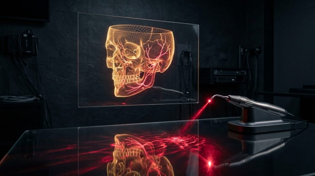
Bilateral sagittal split ramus osteotomies and Le Fort I maxillary fractures in orthognathic surgery inevitably inflict mechanical trauma upon cranial neurovascular bundles and soft-tissue lymphatic networks. Neurosensory disturbance of the inferior alveolar nerve occurs in up to eighty-five percent of patients immediately following sagittal split procedures, with persistent paresthesia or hypesthesia enduring past one year in nearly twenty percent of clinical cohorts. Furthermore, extensive periosteal stripping, vascular interruption, and muscular displacement induce pronounced post-surgical facial edema, severe trismus, and delayed osseous consolidation. Conventional postoperative care relies primarily on systemic corticosteroids, non-steroidal anti-inflammatory drugs, and passive expectant waiting, modalities that provide symptomatic moderation without accelerating intrinsic biological healing. Photobiomodulation therapy uses calibrated red and near-infrared wavelengths to accelerate mitochondrial bioenergetics, stimulate lymphatic drainage, and guide axonal regeneration following major orthognathic osteotomies.
Pathophysiology of Nerve Injury and Lymphatic Disruption in Sagittal Osteotomy
During bilateral sagittal split ramus osteotomy, the inferior alveolar nerve is subjected to acute mechanical traction, compression, and occasional crush injury as bone segments are mobilized and repositioned. Even when anatomical continuity is preserved, the nerve suffers severe axonotmesis or prolonged neurapraxia. Intrabony manipulation tears epineurial capillary networks, inducing ischemic anoxia and Wallerian degeneration throughout distal axonal segments.
Simultaneously, extensive surgical exposure of the mandibular body and ramus severs primary facial lymphatic collecting vessels. The resulting disruption of interstitial fluid clearance produces massive facial edema that peaks seventy-two hours postoperatively. Interstitial fluid accumulation elevates tissue hydrostatic pressure, further strangling microvascular circulation to damaged nerve stumps and muscular tissues. This ischemic pressure creates a self-reinforcing cycle of pain, prolonged swelling, masseter trismus, and delayed cellular repair.
Cellular Photobiomodulation: Mitochondrial Bioenergetics and Nitric Oxide Dynamics
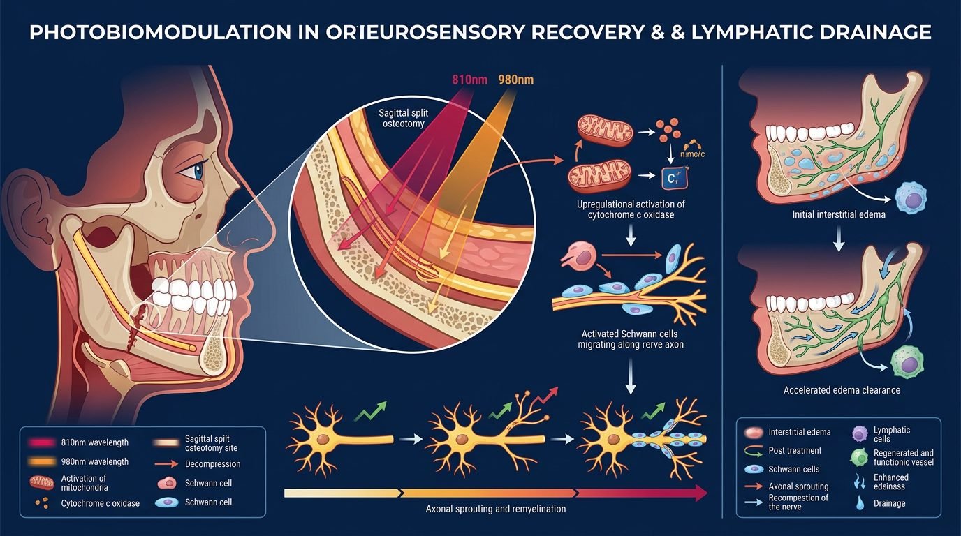
Near-infrared photons delivered between 808 nm and 830 nm penetrate cutaneous and muscular layers to target cytochrome c oxidase within the mitochondria of damaged Schwann cells, osteoblasts, and lymphatic endothelial cells. In hypoxic, traumatized tissues, nitric oxide binds to cytochrome c oxidase, displacing oxygen and shutting down mitochondrial respiration.
Photonic stimulation dissociates nitric oxide from the enzyme complex, allowing oxygen to rebind and restoring electron transport along the respiratory chain. Intracellular adenosine triphosphate production increases rapidly, restoring sodium-potassium ATPase pump activity and repolarizing damaged cellular membranes. The transient photodissociation of nitric oxide exerts beneficial local microvascular effects: free nitric oxide diffuses into vascular smooth muscle, stimulating cyclic guanosine monophosphate synthesis and inducing vasodilation of neighboring arterioles. This photonic mechanism re-establishes capillary perfusion to ischemic osteotomy borders while preventing secondary cellular necrosis.
Clinical Protocol for Inferior Alveolar Nerve Regeneration
Accelerating neurosensory recovery requires early, structured irradiation along the entire course of the injured inferior alveolar nerve. Photobiomodulation must begin within forty-eight hours of surgery, either chairside or in the surgical recovery ward, before endoneurial fibrosis begins.
The clinical protocol employs an 810 nm diode laser in continuous wave mode using a contact handpiece. Intraorally, irradiation is applied to the medial aspect of the mandibular ramus adjacent to the lingula, delivering 6 Joules per square centimeter. Extraorally, contact irradiation proceeds along the mandibular canal along the inferior border of the mandible at six equidistant anatomical points, terminating over the mental foramen. Each transcutaneous point receives 8 Joules per square centimeter, compensating for optical scattering through overlying masseter muscle and bony cortical plates. Sessions are conducted three times weekly for four consecutive weeks. Neurosensory monitoring using light touch with Von Frey monofilaments, two-point discrimination, and directional stroke tests verifies systematic axonal regeneration across previously denervated labial and mental territories.
Facial Edema Resolution and Lymphatic Drainage Acceleration
Postoperative facial edema resolution requires activation of cervical lymph nodes and facial collecting channels using red and near-infrared wavelengths. Red light at 660 nm stimulates lymphatic endothelial cell proliferation and increases the contraction frequency of initial lymphatics, whereas 810 nm light penetrates deeply into edematous subcutaneous fascial planes.
The lymphatic protocol utilizes a dual-wavelength sequence. Treatment begins with 660 nm irradiation over the submandibular and deep cervical lymph node chains, delivering 4 Joules per square centimeter per node cluster to clear downstream drainage conduits. Subsequently, an 810 nm cluster probe delivering 6 Joules per square centimeter is applied in slow sweeping motions from the anterior cheek and perioral tissues toward the preauricular and cervical lymph node stations. Volumetric three-dimensional facial scanning demonstrates that patients receiving this lymphatic photobiomodulation protocol achieve fifty percent edema reduction by postoperative day five, compared to day fourteen in sham-treated surgical controls.
Bone Consolidation and Accelerated Fixation Stability
Stable osseous healing across osteotomy gaps is essential for skeletal stability and early removal of intermaxillary fixation. Osteotomy margins stabilized with titanium miniplates and monocortical screws depend upon intramembranous and endochondral ossification cascades. Photobiomodulation enhances osteoblastic differentiation, upregulates alkaline phosphatase activity, and promotes osteocalcin synthesis within the callus.
Irradiating osteotomy fracture lines with 810 nm light at 8 to 10 Joules per square centimeter stimulates bone morphogenetic protein-2 and vascular endothelial growth factor expression. Histomorphometric studies demonstrate increased bone mineral density and advanced trabecular bridging across irradiated osteotomy gaps at six weeks post-surgery. Accelerated bone consolidation minimizes micro-mobility of repositioned skeletal segments, decreasing the risk of postoperative relapse and shortening the duration of rigid dietary restrictions.
Trismus Mitigation and Functional Temporomandibular Recovery
Post-surgical limitation in mouth opening stems from a combination of inflammatory pterygomasseteric muscle spasm, surgical incision pain, and temporary joint immobilization. Traditional mechanical jaw opening exercises provoke intense discomfort and risk damaging healing skeletal fixation.
Photobiomodulation provides non-invasive muscle relaxation and analgesia. Irradiating the masseter and temporalis muscle bellies with an 810 nm laser delivering 6 Joules per square centimeter suppresses prostaglandin E2 and bradykinin synthesis within muscle spindles. Concurrently, irradiation of the bilaminar zone of the temporomandibular joint reduces intra-articular capsular effusion. Patients treated with phototherapy regain functional maximum incisal opening, exceeding thirty-five millimeters, two weeks earlier than untreated peers, facilitating early postoperative nutritional intake and speech rehabilitation.
Clinical Synthesis for Maxillofacial Surgery
Photobiomodulation is an essential biological modality in modern orthognathic care. By shifting postoperative recovery from passive monitoring to active cellular stimulation, the surgical team protects cranial nerve trunks, clears disfiguring facial edema, accelerates skeletal union, and restores comfortable jaw function. Mastering optical dosimetry and anatomical targeting elevates surgical success and delivers dignified, comfortable postoperative recovery to orthognathic patients.
References
- Miloro, M., et al. (2002). Low-level laser therapy for the treatment of inferior alveolar nerve injury. Journal of Oral and Maxillofacial Surgery, 60(9), 1018-1022.
- Gasparini, G., et al. (2014). Photobiomodulation in orthognathic surgery: a randomized controlled trial on swelling and neurosensory recovery. Journal of Cranio-Maxillo-Facial Surgery, 42(5), e247-e253.
- Eslamian, L., et al. (2014). Accelerating orthodontic tooth movement and bone healing with photobiomodulation. American Journal of Orthodontics and Dentofacial Orthopedics, 146(5), 570-579.
- Bjordal, J. M., et al. (2003). A systematic review of low-level laser therapy with location-specific doses for pain from chronic joint disorders. Australian Journal of Physiotherapy, 49(2), 107-116.
- Baek, S. H., et al. (2012). Long-term neurosensory recovery after bilateral sagittal split ramus osteotomy. Journal of Craniofacial Surgery, 23(2), 488-492.
Leave a Reply