
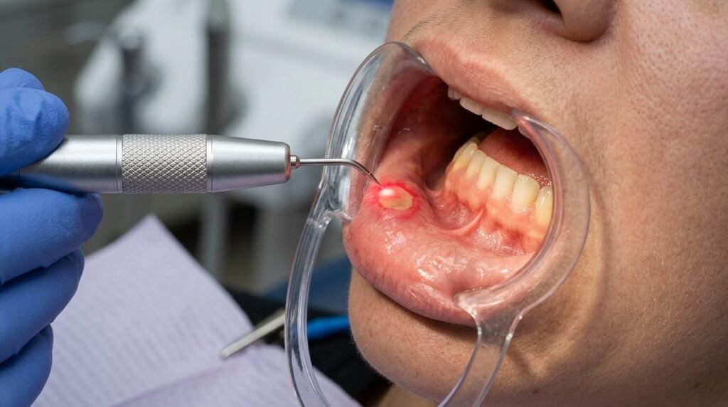
Recurrent ulcerative oral lesions, notably recurrent aphthous stomatitis and herpes simplex labialis, impose substantial functional morbidity on affected individuals. Mastication, speech articulation, and oral hygiene maintenance become severely compromised due to intense localized nociceptor activation. Conventional pharmacotherapies, including topical corticosteroids, barrier pastes, and systemic antivirals, provide modest clinical improvements but often fail to accelerate mucosal re-epithelialization or eliminate recurrence cycles. Photobiomodulation therapy offers a non-thermal, non-pharmacologic intervention capable of alleviating acute neuropathic discomfort immediately while accelerating cellular tissue regeneration. Red (630 to 660 nm) and near-infrared (810 to 830 nm) low-level laser irradiation interacts with respiratory chromophores in compromised mucosal cells. This biostimulative cascade accelerates fibroblastic proliferation, modulates pro-inflammatory cytokines, suppresses peripheral nociceptive transmission, and extends disease-free remission intervals without inducing adverse systemic reactions.
Pathophysiology of Mucosal Ulceration and Herpes Latency
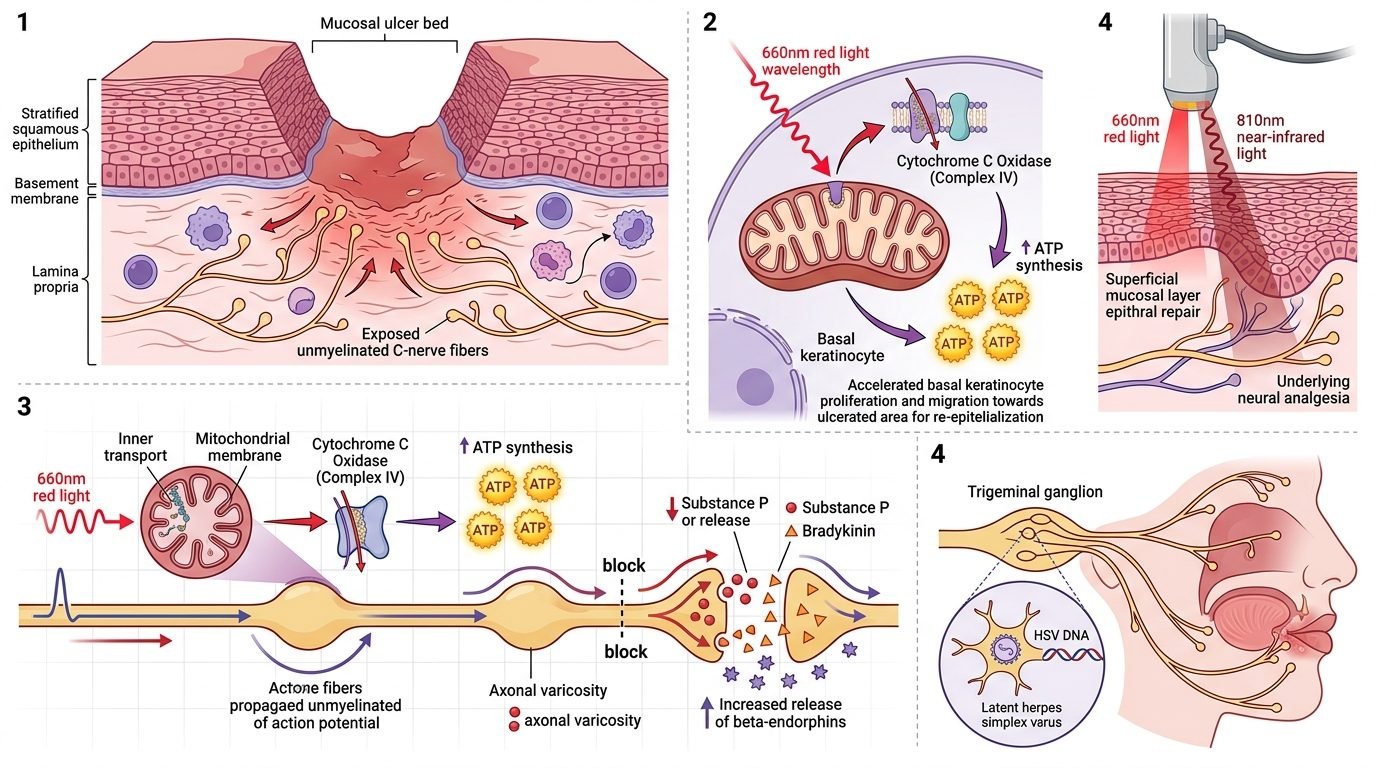
Recurrent aphthous stomatitis represents a cell-mediated immune dysregulation characterized by localized T-lymphocyte cytotoxicity against oral mucosal keratinocytes. Interleukin-2, interferon-gamma, and tumor necrosis factor-alpha drive focal epithelial necrosis, producing painful fibrinous pseudomembranous craters surrounded by erythematous borders. In contrast, herpes simplex virus type one establishes lifelong latent infection within the trigeminal sensory ganglion. Reactivation triggered by psychological stress, ultraviolet radiation, or local mechanical trauma causes viral particles to travel down peripheral axons to the perioral vermilion border. Intraepithelial ballooning, acantholysis, and vesicular eruption follow, terminating in painful crusting lesions.
Both pathologies share a common clinical hurdle: extreme focal pain driven by exposed unmyelinated nociceptive free nerve endings. Traditional therapeutic approaches act topically to coat or numb the ulcer bed temporarily. However, chemical analgesics do not stimulate tissue repair at the cellular level. Photobiomodulation bypasses surface barriers, delivering photons that penetrate through the fibrinous exudate to interact directly with basal keratinocytes, fibroblasts, endothelial cells, and peripheral nerve endings.
Mitochondrial Photoreception and Cellular Re-Epithelialization
The biological acceleration of mucosal healing begins within unit four of the mitochondrial respiratory chain: cytochrome c oxidase. Low-level laser irradiation in the 660 nm red spectrum matches the electronic absorption bands of oxidized copper centers within the enzyme. Photonic absorption promotes the catalytic dissociation of inhibitory nitric oxide, increasing the mitochondrial proton motive force. Oxygen consumption accelerates, driving significant upregulation of adenosine triphosphate synthesis. Cellular energy reserves replenish rapidly, empowering dormant basal keratinocytes at ulcer margins to initiate migratory and proliferative cycles.
Concurrently, photobiomodulation modulates the local inflammatory microenvironment. Low-level laser irradiation downregulates gene expression of tumor necrosis factor-alpha, interleukin-1 beta, and cyclooxygenase-2 while upregulating transforming growth factor-beta and basic fibroblast growth factor. This cytokine shift curtails destructive neutrophilic infiltration and recruits reparative M2 macrophages to the wound bed. Fibroblasts synthesize organized type one and type three collagen fibrils, establishing a healthy granulation tissue base. Clinical trials confirm that laser-treated aphthous ulcers exhibit complete re-epithelialization within three to four days, compared to ten to fourteen days in untreated natural healing cycles.
Electrophysiological Nociceptive Inhibition and Immediate Analgesia
One of the most remarkable clinical attributes of photobiomodulation is the immediate reduction in acute ulcer pain. Patients consistently report substantial analgesic relief within minutes of laser application, allowing comfortable mastication and speech. This immediate analgesia operates through direct electrophysiological effects on peripheral nerve fibers. High-fluence near-infrared photons (810 to 830 nm) temporarily reduce mitochondrial membrane potentials in unmyelinated C-fibers, inducing axonal varicosities that block fast action potential conduction.
Additionally, laser irradiation alters the biochemical milieu surrounding peripheral nociceptors. Photobiomodulation suppresses the local release of bradykinin, histamine, and substance P from degranulating mast cells and primary sensory terminals. Local tissue acidosis neutralizes as capillary perfusion clears acidic metabolic byproducts. Furthermore, low-level laser exposure stimulates the local release of endogenous opioids, particularly beta-endorphins, within the mucosal connective tissue. This multi-target neural inhibition dampens central pain sensitization, providing durable analgesic relief that outlasts the duration of the laser session.
Intervention Protocols for Herpes Simplex Labialis
The clinical efficacy of photobiomodulation in herpes labialis depends critically on the stage of viral reactivation at the time of intervention. Applying laser therapy during the prodromal phase, characterized by localized itching, tingling, or erythema before vesicle appearance, can abort vesicle development entirely. Photons delivered to the perioral tissue stimulate localized microcirculation and recruit cytotoxic T-lymphocytes, boosting host immune defenses before intraepithelial viral replication causes extensive tissue destruction.
When patients present with established vesicles or ruptured crusts, photobiomodulation accelerates crust shedding and minimizes secondary bacterial superinfections. A dual-wavelength approach yields optimal outcomes: an initial pass of 660 nm red light (2 to 4 Joules per square centimeter) stimulates superficial epithelial re-epithelialization, followed by an 810 nm near-infrared pass (4 to 6 Joules per square centimeter) targeting deep dermal and neural structures. Longitudinal observational studies demonstrate that recurrent herpes patients receiving repeated photobiomodulation regimens experience substantial reductions in recurrence frequency, with remission periods extending from months to several years.
Clinical Dosimetry and Non-Contact Delivery Protocols
Successful management of oral mucosal ulcerations demands precise non-contact laser dosimetry. Direct contact with active herpetic vesicles or infected aphthous craters must be avoided to prevent cross-contamination and mechanical pain. The laser handpiece should be held perpendicularly at a distance of two to four millimeters from the lesion surface. The beam sweeps slowly across the entire ulcer bed, including a two-millimeter halo of surrounding erythematous tissue.
Therapeutic energy densities should range between 3 and 6 Joules per square centimeter per point, delivered in continuous wave mode with power settings maintained between 50 and 100 milliwatts. Low power settings eliminate any risk of photothermal sensation, ensuring the treatment remains entirely painless without local anesthesia. For aphthous stomatitis, two to three sessions spaced 24 to 48 hours apart achieve complete clinical resolution. Recording accurate physical parameters guarantees predictable, reproducible clinical success across diverse patient populations.
Clinical Significance in Humanized Oral Care
Integrating photobiomodulation into mucosal pathology management transforms the patient clinical experience and treatment trajectory. Patients suffering from debilitating oral ulcerations frequently endure chronic discomfort due to the limited efficacy of conventional pharmacological ointments. Offering immediate, non-invasive pain relief without pharmaceutical toxicity strengthens the therapeutic bond between practitioner and patient. Dental teams equipped with biophotonic expertise deliver compassionate, biologically sound interventions that restore oral comfort rapidly and predictably.
References
- Albrektson, M., et al. (2014). Photobiomodulation therapy in recurrent aphthous stomatitis: A systematic review and meta-analysis. Lasers in Medical Science, 29(4), 1475-1483.
- Bello-Silva, M. S., et al. (2010). Low-level laser therapy for herpes simplex labialis: A randomized controlled clinical trial. Photomedicine and Laser Surgery, 28(5), 651-656.
- Hamblin, M. R. (2017). Mechanisms and applications of the anti-inflammatory effects of photobiomodulation. AIMS Biophysics, 4(3), 337-361.
- de Souza, F. T., et al. (2010). Laser therapy for recurrent aphthous stomatitis: an open, prospective study. Journal of Oral Pathology & Medicine, 39(8), 643-647.
- Karu, T. I. (2008). Mitochondrial mechanisms of photobiomodulation in tissue repair. Photomedicine and Laser Surgery, 26(3), 193-200.
Leave a Reply