

Primary Sjögren’s syndrome is a chronic systemic autoimmune disorder characterized by progressive lymphocytic infiltration of exocrine glands, resulting in profound xerostomia, keratoconjunctivitis sicca, and catastrophic degradation of oral health. Autoreactive CD4-positive T-helper cells and B-lymphocytes organize into ectopic germinal centers within the parotid and submandibular glands, triggering apoptotic destruction of secretory acinar epithelial cells. The resulting severe salivary hypofunction causes rampant cervical caries, persistent oral candidiasis, dysphagia, dysgeusia, and mucosal ulcerations. Systemic parasympathomimetic sialogogues such as pilocarpine and cevimeline provide limited, transient symptomatic benefit while producing intolerable cholinergic side effects, including profuse diaphoresis, nausea, and urinary frequency. Photobiomodulation (PBM) utilizing near-infrared laser radiation (808 nm) provides a targeted, non-invasive biophysical intervention capable of stimulating residual glandular parenchyma, enhancing microvascular perfusion, and attenuating intra-glandular inflammatory cytokine cascades.
The cellular bioenergetics underlying photobiomodulation in damaged salivary parenchyma depend directly on targeted mitochondrial photo-reception. Photons emitted at the near-infrared wavelength of 808 nm possess high optical penetration depth through overlying subcutaneous adipose, platysma, and masseteric muscular tissues, efficiently reaching the deep lobes of the parotid, submandibular, and sublingual glands. At the subcellular level, near-infrared radiation is selectively absorbed by cytochrome c oxidase (complex IV) located within the inner mitochondrial membrane of residual acinar cells, intercalated ductal cells, and striated ductal epithelium. This photonic absorption accelerates electron transport chain kinetics, elevating intracellular adenosine triphosphate (ATP) synthesis and modulating physiological levels of reactive oxygen species (ROS) that act as essential secondary messengers. Intracellular ATP accumulation directly powers the active basolateral sodium-potassium-chloride cotransporter (NKCC1) and the primary sodium-potassium ATPase pumps, while simultaneously facilitating intracellular calcium ion mobilization to open apical calcium-activated chloride channels (TMEM16A/ANO1). This coordinated ionic flux drives the transepithelial osmotic gradient required for primary isotonic fluid secretion into the glandular acinar lumina.
Anti-inflammatory immunomodulation represents a primary therapeutic mechanism in autoimmune sialadenitis. The chronic destruction of salivary tissue is driven by high concentrations of pro-inflammatory cytokines, specifically tumor necrosis factor-alpha (TNF-alpha), interleukin-1-beta (IL-1-beta), interleukin-6 (IL-6), and interferon-gamma (IFN-gamma), which sustain acinar apoptosis and inhibit neural acetylcholine release. Laser irradiation at 808 nm downregulates the nuclear factor kappa-light-chain-enhancer of activated B cells (NF-kB) signaling pathway within glandular parenchymal cells and infiltrating mononuclear leukocytes. Longitudinal clinical evaluations demonstrate measurable reductions in salivary and serum concentrations of TNF-alpha and IL-6 following protocol-driven PBM therapy. Concurrently, laser irradiation upregulates transforming growth factor-beta (TGF-beta) and interleukin-10 (IL-10), fostering a local tolerogenic microenvironment that curtails autoantibody-mediated cytotoxicity.
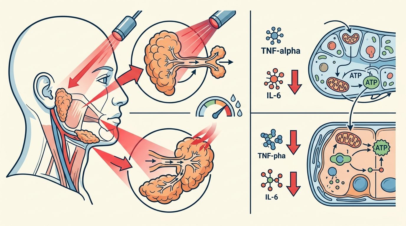
Microvascular revascularization and tissue perfusion constitute another vital dimension of glandular rehabilitation. Autoimmune infiltration induces progressive microvascular obliteration and parenchymal ischemia within salivary lobules. Near-infrared photobiomodulation triggers endothelial nitric oxide synthase (eNOS) activation, producing localized nitric oxide release that induces arteriolar vasodilation. Enhanced microvascular blood flow increases oxygenation and nutrient delivery to remaining viable acini. Furthermore, sustained PBM protocols stimulate endothelial cell proliferation and upregulate vascular endothelial growth factor (VEGF), promoting functional neoangiogenesis in chronically ischemic glandular zones and reversing cellular atrophy in borderline-viable acinar units.
Quantifiable salivary secretory outcomes demonstrate clinical efficacy. Standardized sialometry protocols measuring unstimulated whole salivary flow (UWSF) and stimulated whole salivary flow (SWSF) demonstrate statistically significant and clinically meaningful improvements following comprehensive PBM regimens. In randomized controlled clinical trials, patients receiving active 808 nm laser therapy exhibited a forty to seventy-five percent increase in baseline unstimulated salivary flow rates after six to ten treatment sessions, whereas sham-irradiated controls showed negligible changes. Subjective xerostomia severity, assessed using the validated Xerostomia Inventory (XI) and visual analogue scales (VAS), demonstrated durable reductions in oral dryness, nocturnal awakening, and difficulty swallowing dry foods, with benefits persisting for up to twelve to sixteen weeks post-treatment.
Salivary qualitative composition and mucosal defense undergo positive biochemical remodeling. Beyond increasing raw fluid volume, photobiomodulation alters the organic composition of secreted saliva. Laboratory salivary proteomic analyses reveal elevated concentrations of secretory immunoglobulin A (sIgA), alpha-amylase, lysozyme, and lactoferrin following laser protocols. Restoring these antimicrobial proteins and buffering bicarbonates re-establishes intraoral ecological stability, reducing carriage rates of Candida albicans and mutans streptococci. Clinical examinations document a significant decline in secondary angular cheilitis, oral candidiasis relapses, and painful mucosal erythema.
Dosimetric parameters and clinical application geometry must be strictly standardized to achieve therapeutic energy thresholds within dense, fibrotic glandular tissue. Treatment is delivered using an 808 nm continuous-wave gallium-aluminum-arsenide (GaAlAs) semiconductor diode laser equipped with a calibrated flat-top beam profile handpiece or a divergent fiber-optic cluster probe to ensure uniform photonic distribution without focal hotspots. For the anatomical volume of the parotid gland, transcutaneous irradiation is applied across eight to ten distinct anatomical points distributed methodically over the superficial and deep lobes, delivering 4 to 6 joules per square centimeter per point with an irradiance maintained at one hundred milliwatts per square centimeter and a spot size of 0.8 square centimeters. For the submandibular gland, four to six discrete points along the submandibular triangle are irradiated using identical dosimetric parameters. The total cumulative energy delivered per session ranges from twenty-four to thirty-six joules per parotid gland and twelve to eighteen joules per submandibular gland. Treatments are administered twice weekly across six to eight consecutive weeks, followed by monthly maintenance sessions to sustain long-term glandular cellular bioenergetics.
Diagnostic screening and prognostic stratification dictate appropriate patient selection. Clinicians must recognize that photobiomodulation requires viable residual acinar tissue to achieve secretory stimulation. Patients in end-stage Sjögren’s syndrome, where glandular parenchyma has undergone complete fibrous or fatty involution (demonstrated by high-grade parenchymal heterogeneity on salivary ultrasonography or severe atrophy on magnetic resonance imaging), show minimal functional recovery following laser therapy. Conversely, patients with early-to-moderate disease stage, characterized by mild-to-moderate ultrasonographic glandular changes and preserved baseline stimulated flow, demonstrate the most pronounced and durable therapeutic responses.
Safety precautions and oncological contraindications must be strictly observed. Sjögren’s syndrome carries a well-established six-to-twenty-fold increased risk of developing B-cell non-Hodgkin lymphoma, predominantly mucosa-associated lymphoid tissue (MALT) lymphoma of the parotid gland. The clinician must conduct thorough palpation of all major salivary glands and cervical lymph node chains prior to initiating laser therapy. Any persistent, firm, nodular, or asymmetric glandular enlargement mandates immediate diagnostic ultrasonography and biopsy to exclude lymphoid malignancy before applying biostimulative laser energy.
Regarding clinical practice, incorporating near-infrared photobiomodulation into the comprehensive multidisciplinary management of Sjögren’s syndrome offers a highly effective, biologically sound modality that transcends purely palliative topical lubricating sprays and poorly tolerated systemic cholinergic pharmaceuticals. By systematically boosting mitochondrial bioenergetics, mitigating chronic intra-glandular pro-inflammatory cytokine loops, enhancing microvascular capillary perfusion, and stimulating residual acinar secretory activity, 808 nm diode laser therapy restores endogenous whole saliva production. Clinicians adopting structured, reproducible dosimetric protocols can deliver substantial, long-term symptomatic relief, safeguard natural dentitions, and prevent catastrophic oral health breakdown in this vulnerable autoimmune patient population.
Leave a Reply