

Photobiomodulation exerts measurable neuroprotective and inhibitory actions across peripheral trigeminal branches by modulating axoplasmic flow, suppressing nociceptive action potentials, and decreasing neurogenic inflammation. Trigeminal neuralgia (TN) and persistent idiopathic dentoalveolar pain (PIDAP, formerly atypical odontalgia) represent some of the most agonizing neuropathic conditions encountered in oral healthcare. Classical trigeminal neuralgia presents as paroxysmal, lancinating, electric shock-like spasms triggered by light tactile stimuli within the distribution of the maxillary (V2) or mandibular (V3) divisions. Conversely, persistent dentoalveolar pain manifests as continuous, unremitting, burning or throbbing pain localized to a dentoalveolar site following endodontic treatment or extraction, absent identifiable pathology. Misdiagnosis frequently drives clinicians to perform destructive, irreversible dental procedures, including repeat endodontic retreatments, apicoectomies, and extractions, which amplify central sensitization. Applying non-invasive photobiomodulation provides conservative neurosensory modulation while protecting intact dentoalveolar architecture.
Diagnostic Differentiation: Classical Neuralgia versus Dentoalveolar Neuropathy
Rigorous clinical differentiation between classical neurovascular compression and peripheral neuropathic pain determines therapeutic feasibility. Classical trigeminal neuralgia arises primarily from microvascular pulsatile compression of the trigeminal sensory nerve root at the prepontine root entry zone, most commonly by an aberrant loop of the superior cerebellar artery. This neurovascular conflict induces focal focal demyelination, ectopic axonal pacemakers, and cross-excitation (ephaptic transmission) between tactile A-beta fibers and nociceptive A-delta and C-fibers.
In contrast, persistent idiopathic dentoalveolar pain reflects peripheral neuroplastic remodeling following minor surgical trauma to pulpal or periodontal terminal nerve endings. The diagnostic criteria defined by the International Classification of Orofacial Pain (ICOP) require deep aching pain persisting daily for more than two hours per day for at least three months, without anatomical, radiographical, or endodontic abnormalities. Photobiomodulation cannot correct mechanical arterial loops inside the posterior cranial fossa; in classical TN, its role remains palliative and adjuvant to anticonvulsants like carbamazepine or oxcarbazepine. However, in peripheral post-traumatic trigeminal neuropathies and PIDAP, PBM acts directly upon sensitized peripheral nerve terminals, modifying the hyperactive peripheral drive feeding dorsal horn sensitization.
Neurobiological Mechanisms: Conduction Block and Axoplasmic Varicosities
The biophysical foundation of laser analgesia in neuropathic pain operating under high irradiance involves fast axonal conduction inhibition. When peripheral nerve trunks receive continuous-wave near-infrared laser energy (810 nm to 830 nm) at irradiances exceeding 300 mW/cm2, photon absorption by mitochondrial chromophores produces a transient elevation in intracellular adenosine triphosphate (ATP) and localized hyperpolarization.
Higher energy densities induce reversible varicosities within unmyelinated nociceptive axons. This physiological varicosity formation temporarily interrupts microtubule-based fast axonal transport, slowing saltatory propagation along A-delta and C-fibers. Electrophysiological recordings demonstrate a significant reduction in compound action potential amplitude and an increased latency of nociceptive impulses within fifteen minutes of irradiation. Furthermore, PBM suppresses the local secretion of pro-inflammatory neuropeptides, specifically substance P and calcitonin gene-related peptide (CGRP), from peripheral terminal boutons. Attenuating neuropeptide extravasation eliminates neurogenic plasma extravasation and blunts peripheral mechanoreceptor hypersensitivity.
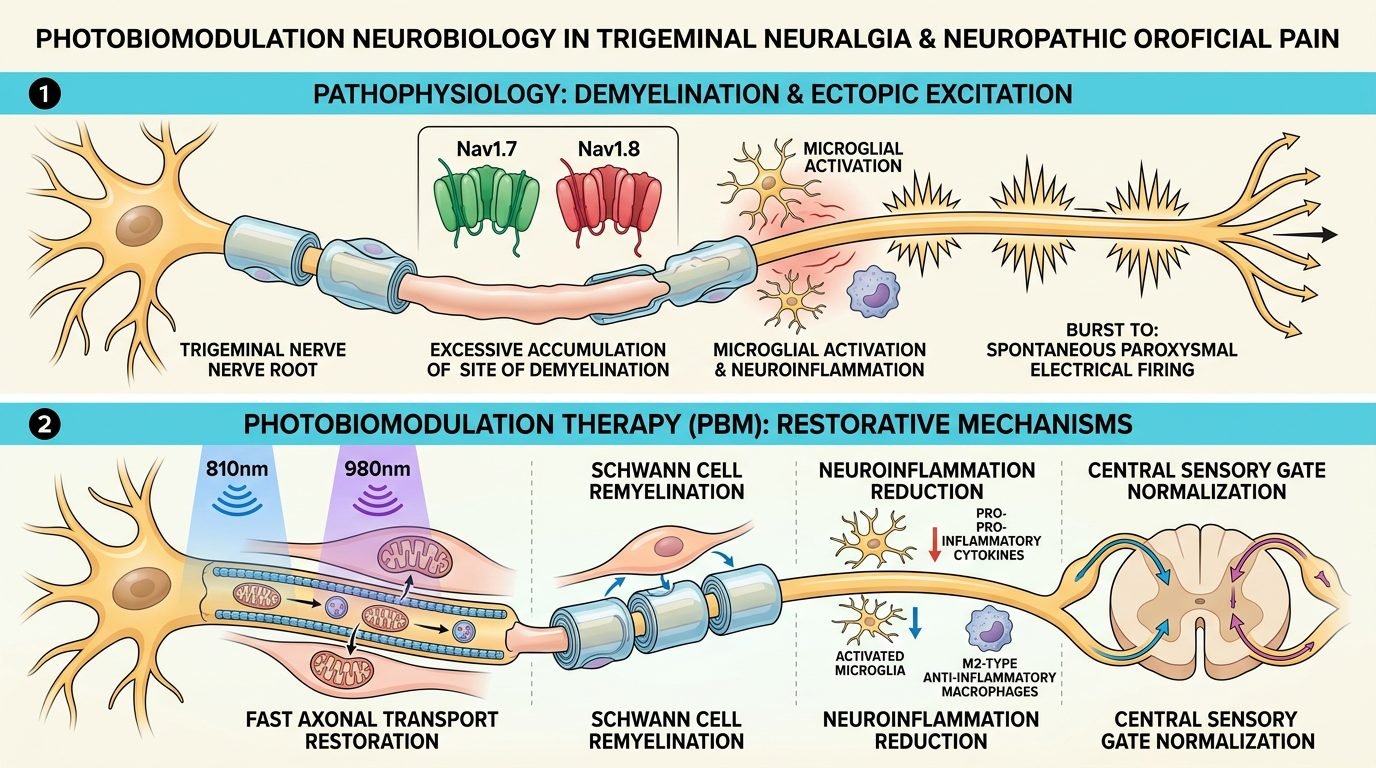
At the dorsal root ganglion and trigeminal nucleus caudalis level, this peripheral silencing exerts descending modulatory influences. Animal and clinical biomarker assays show that localized near-infrared irradiation downregulates pro-inflammatory cytokines, specifically tumor necrosis factor-alpha (TNF-alpha) and interleukin-1 beta (IL-1beta), within perineural Schwann cells. Simultaneously, systemic endogenous opioid peptide release, including beta-endorphin, increases in cerebrospinal fluid, elevating central nociceptive thresholds.
Extraoral and Intraoral Irradiation Protocols
Delivering sufficient photon energy to affected trigeminal branches requires combined intraoral and extraoral irradiation geometries. Because cortical bone and overlying facial soft tissues attenuate near-infrared light by 80% to 90%, clinicians must utilize wavelengths with superior tissue penetration depths, specifically 808 nm to 830 nm, or dual-wavelength combinations incorporating 904 nm superpulsed diodes.
For maxillary branch involvement (V2), extraoral irradiation targets the infraorbital foramen (located approximately 5 to 8 mm below the infraorbital rim) and the pre-auricular fossa anterior to the tragus. The probe is applied in firm perpendicular contact with the skin: wavelength 808 nm, power 200 mW, continuous wave, spot size 0.5 cm2, energy density 8 J/cm2 per point, duration 60 seconds per point. Intraorally, irradiation is directed along the canine fossa and the greater palatine foramen, delivering 4 J/cm2 per mucosal point. For mandibular involvement (V3), the extraoral protocol targets the mental foramen (between mandibular premolar apices) and the mandibular notch overlying the infratemporal fossa, delivering 8 to 10 J/cm2. Intraorally, the lingual aspect of the retromolar trigone is irradiated adjacent to the lingual and inferior alveolar nerve entrance, applying 6 J/cm2.
Dosimetric Precision and the Biphasic Dose-Response
Neuropathic pain protocols are strictly governed by the Arndt-Schultz biphasic dose-response curve. Insufficient photonic energy (less than 1 J/cm2 per site) fails to activate mitochondrial cytochrome c oxidase and elicits negligible analgesic response. Conversely, excessive radiant exposure (greater than 16 J/cm2 per point) or excessive power causing thermal elevation above 42 degrees Celsius produces paradoxical nociceptive stimulation, exacerbating paroxysmal shooting pains.
Clinicians must avoid moving the handpiece continuously in high-speed scanning motions, as dynamic painting dilutes energy density and fails to achieve the photon threshold required for neural conduction slowing. Beam divergence across facial adipose tissue must be accounted for by maintaining perpendicular contact pressure with the cutaneous boundary. Treatment sessions should be scheduled three times weekly for the first two weeks, tapering to twice weekly for four subsequent weeks. Total energy per anatomical quadrant should be constrained to 30 to 50 Joules per session to prevent neuroinflammatory rebounds.
Clinical Outcomes and Adjunctive Pharmaceutical Management
Clinical trials investigating photobiomodulation in trigeminal neuralgia demonstrate substantial reductions in visual analog scale (VAS) pain ratings, with over 60% of patients achieving greater than 50% symptom reduction. More significantly, PBM facilitates downward titration of carbamazepine, gabapentin, or pregabalin, mitigating disabling pharmacological side effects such as somnolence, cognitive blunting, and hepatic strain.
In persistent dentoalveolar pain, laser therapy diminishes allodynia to toothbrushing and relieves burning sensations in gingival tissues. Nevertheless, clinicians must maintain realistic expectations. If a patient with presumed TN demonstrates progressive sensory deficits, corneal numbness, or motor weakness, urgent neurosurgical imaging via high-resolution 3D FIESTA MRI is mandatory to rule out cerebellopontine angle tumors or multiple sclerosis plaques. Photobiomodulation serves as a sophisticated biophysical intervention within a comprehensive multidisciplinary pain strategy.
References
- Eslamian, L., et al. (2014). The effect of low-level laser therapy on sensory recovery of the inferior alveolar nerve after bilateral sagittal split osteotomy: a double-blind, split-mouth, randomized clinical trial. Journal of Oral and Maxillofacial Surgery, 72(7), 1411-1419.
- Falaki, F., et al. (2014). Management of facial pain in patients with trigeminal neuralgia using low-level laser therapy: a randomized controlled trial. Neurosciences (Riyadh), 19(4), 310-314.
- International Headache Society. (2020). International Classification of Orofacial Pain, 1st edition (ICOP). Cephalalgia, 40(2), 129-221.
- Peñarrocha-Diago, M., et al. (2012). Post-traumatic trigeminal neuropathic pain: review of current concepts and treatment modalities. Medicina Oral, Patologia Oral y Cirugia Bucal, 17(5), e743-e748.
- Svensson, P., et al. (2011). Chronic oral pain: etiology, diagnosis, and treatment. Dental Clinics of North America, 55(1), 105-121.
Leave a Reply