
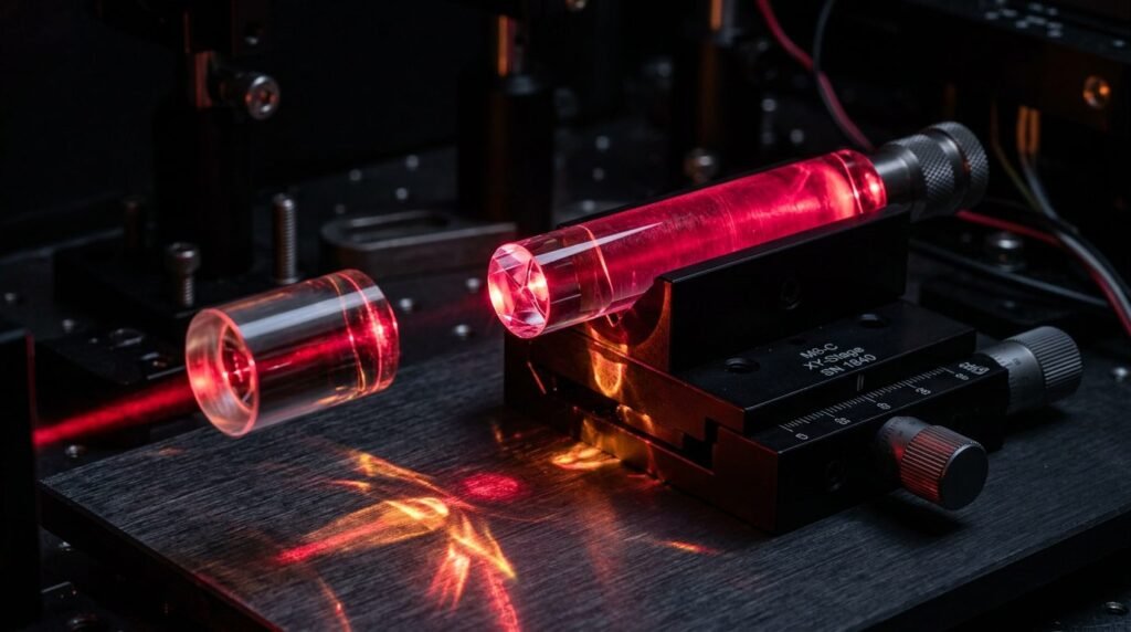
Pulsed Nd:YAG laser irradiation at 1,064 nm selectively ablates diseased pocket epithelium while stimulating true periodontal regeneration characterized by new cementum, periodontal ligament, and alveolar bone. Conventional open-flap periodontal surgery resects soft tissues and creates gingival recession and root exposure. Mechanical curettage lacks optical selectivity, sacrificing healthy connective fibers. The free-running pulsed Nd:YAG laser operates at a wavelength where melanin and hemoglobin exhibit high absorption, whereas water and dental hard tissues exhibit minimal optical attenuation. This physical characteristic enables the laser beam to pass harmlessly through root dentin while selectively vaporizing pigmented periodontal pathogens and chronified pocket lining. Human histological studies demonstrate that the Laser-Assisted New Attachment Procedure (LANAP) achieves true periodontal regeneration without foreign barrier membranes or synthetic bone grafts.
Optical Physics and Differential Chromophore Absorption at 1,064 nm
The neodymium-doped yttrium aluminum garnet (Nd:YAG) laser emits infrared photons at 1,064 nm. In this near-infrared region, absorption by water and biological hydroxyapatite is negligible. Consequently, dry enamel and cementum remain unaffected by transmitted photons. However, the extinction coefficient for dark endogenous chromophores, particularly deoxyhemoglobin and melanin, remains significant.
Periodontal pocket lining affected by chronic inflammatory periodontitis is densely vascularized and infiltrated with pigmented anaerobic bacteria, including Porphyromonas gingivalis. When pulsed 1,064 nm photons enter the pocket, light energy is absorbed selectively by pigmented bacterial membranes and engorged microcapillaries. Free-running pulsed emission utilizes pulse durations of one hundred to one hundred and fifty microseconds with peak powers exceeding one thousand watts. These microsecond bursts vaporize inflamed pocket epithelium and deep granulation tissue. Meanwhile, the thermal relaxation time of adjacent collagenous lamina propria prevents heat accumulation, preserving healthy connective tissue fibers inserted into the root surface.
The LANAP Protocol: Step-by-Step Surgical Bio-Engineering
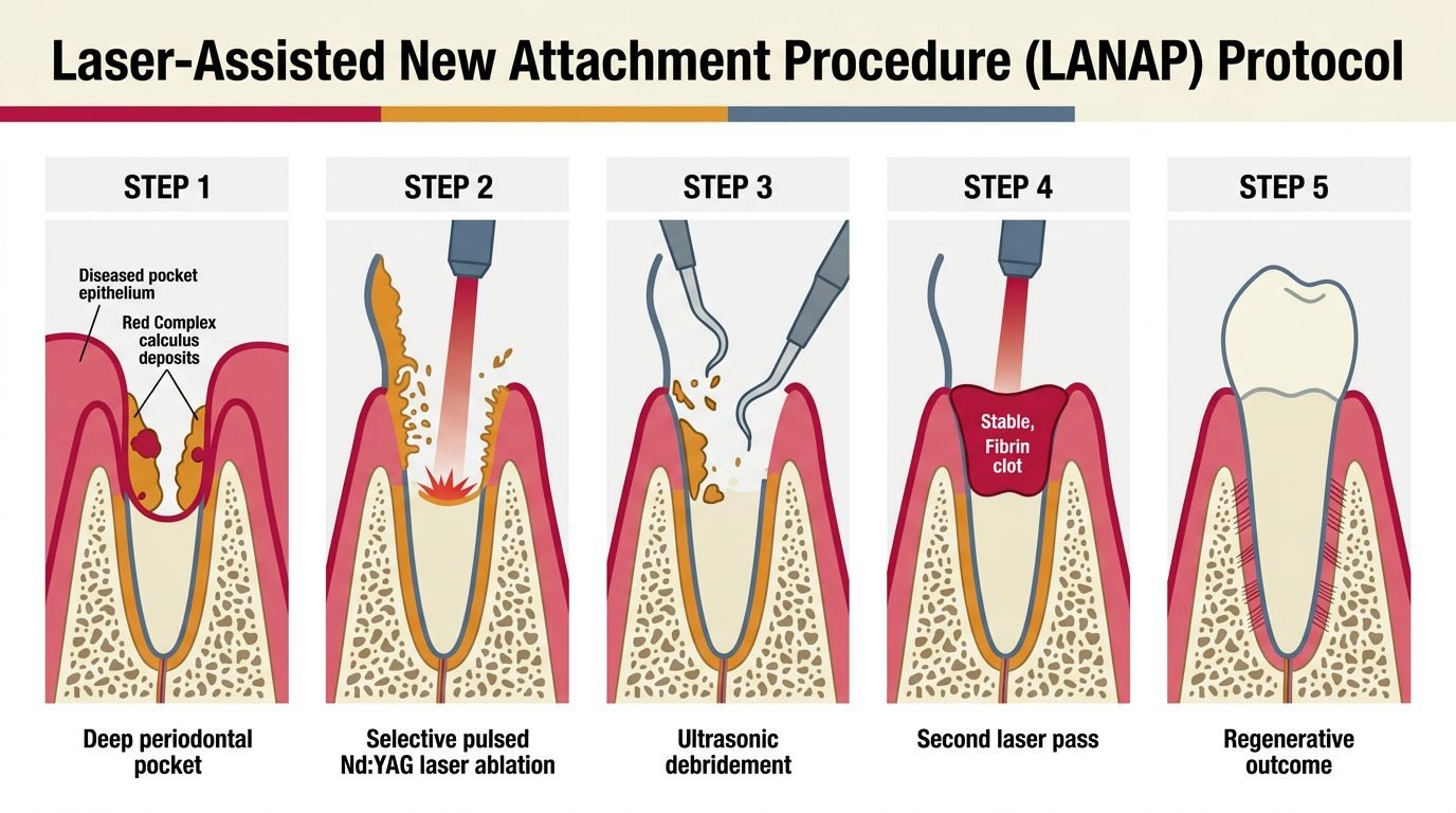
The Laser-Assisted New Attachment Procedure is an evidence-based surgical sequence designed to foster cementum-mediated new attachment. LANAP relies upon two distinct laser passes separated by ultrasonic debridement.
The initial laser pass utilizes a three-hundred-micrometer optical fiber introduced parallel to the root surface down to the osseous defect base. Operating at 3.6 to 4.0 watts, 20 Hertz, and a short pulse duration of one hundred microseconds, the fiber is swept circumferentially. This initial pass ablates diseased sulcular epithelium, lyses dark anaerobes, and denatures subgingival calculus matrices. Calculus softened by photonic absorption becomes porous, significantly easing subsequent removal. The clinician then employs specialized piezoelectric ultrasonic scalers to dislodge root deposits and modify the root morphology without stripping healthy cementum.
The second laser pass alters dosimetry to achieve hemostasis and establish a stable fibrin clot. Operating at higher total energy and long pulse durations around six hundred microseconds, the fiber is swept upward from the defect floor. This pass induces thermal coagulation of deep arterioles and activates fibrinogen into a dense, resilient thermal fibrin clot. The resulting biologic seal adheres tenaciously to the root surface, closing the pocket orifice and preventing down-growth of oral junctional epithelium.
Selective Epithelial Debridement and Connective Tissue Preservation
The barrier to true regeneration after debridement is rapid downgrowth of junctional epithelium, which migrates ten times faster than mesenchymal stem cells, forming non-functional scar attachments.
Pulsed Nd:YAG irradiation circumvents this limitation through optical selectivity. The 1,064 nm energy targets the highly vascularized, inflamed pocket lining, vaporizing ulcerated epithelium down to the basement membrane while sparing deeper collagen bundles. Intact collagen fibers remaining on the root face serve as natural biological scaffolds for migrating periodontal ligament fibroblasts. Histological sections confirm that LANAP-treated pockets show complete de-epithelialization of the sulcular wall, creating a biological window for osteoprogenitor and cementoblast differentiation from the intact periodontal ligament space.
True Periodontal Regeneration Versus Repair: Human Histological Evidence
In periodontology, repair denotes healing by tissue replacement that does not replicate original architecture, exemplified by long junctional epithelium and fibrous scarring. True regeneration requires the tripartite formation of new cellular cementum, functional periodontal ligament fibers oriented perpendicularly to the root, and new alveolar bone.
Human histological biopsies from teeth scheduled for extraction following LANAP therapy provide definitive evidence of true regeneration. Histological evaluations by Nevins and colleagues demonstrated newly formed cementum inserting Sharpey’s fibers into adjacent regenerated alveolar bone in eighty-four percent of treated defects. Regeneration occurred without root resorption or ankylosis. Control teeth treated with conventional scaling and root planing healed uniformly with long junctional epithelium. The presence of a stable, laser-induced fibrin clot prevents junctional epithelial downgrowth, allowing pluripotential mesenchymal cells to orchestrate tissue regeneration.
Occlusal Adjustment and Splinting: Mechanical Stabilization
Periodontal regeneration requires absolute mechanical stability during early healing phases. Micromotion of healing teeth disrupts fragile fibrin clots and shears newly forming capillaries, converting osteogenesis into fibrous scar formation.
The LANAP protocol mandates comprehensive occlusal adjustment immediately preceding or following laser delivery. The clinician eliminates all lateral and protrusive interferences, reducing fremitus to zero. In cases presenting moderate to severe tooth mobility (Miller Grade II or III), extracoronal composite and wire splints must be placed to stabilize the dentition. The patient is instructed to adhere to a soft liquid diet for seven days, avoiding all mechanical brushing or probing of treated pockets for six months post-surgery. Probing immature healing sites prematurely shears nascent Sharpey’s fibers, reversing regenerative cascades.
Comparative Outcomes: LANAP Versus Conventional Resective Flap Surgery
Long-term prospective clinical trials comparing LANAP against modified Widman flap surgery confirm equivalent or superior probing depth reductions and attachment gains. LANAP achieves probing depth reductions between 3.5 and 5.0 millimeters in deep vertical defects while minimizing post-surgical gingival recession.
Patients treated with LANAP experience significantly less postoperative pain, reduced analgesic consumption, and faster resumption of daily activities. The absence of scalpel incisions and vertical releasing cuts eliminates visible tissue shrinkage in aesthetic zones. In patients with complex systemic comorbidities, such as those on anticoagulant therapy or suffering from diabetes, LANAP provides a minimally invasive alternative that achieves infection control and bone regeneration without flap reflection.
Practical Synthesis for the Periodontal Clinician
The pulsed Nd:YAG laser represents a regenerative surgical instrument governed by optical physics. By exploiting differential absorption between vascularized inflammatory tissue and mineralized tooth structures, LANAP eliminates the destructive trauma of resective surgery. Clinicians who follow standardized dosimetry, respect occlusal stability, and protect the healing fibrin seal empower the periodontium to achieve authentic histological regeneration, preserving natural dentition previously deemed hopeless.
References
- Yukna, R. A., et al. (2007). Evaluation of a Nd:YAG laser-assisted new attachment procedure (LANAP) in humans: a histological study. Journal of Periodontology, 78(11), 2110-2122.
- Nevins, M. L., et al. (2012). Human histologic evaluation of periodontal regeneration following the LANAP protocol: a case series. International Journal of Periodontics & Restorative Dentistry, 32(5), 497-507.
- Cobb, C. M. (2006). Lasers in periodontics: a review of the literature. Journal of Periodontology, 77(4), 545-564.
- Harris, D. M., et al. (2004). Laser-assisted new attachment procedure (LANAP): a clinical and histologic overview. General Dentistry, 52(5), 442-449.
- Qadri, T., et al. (2015). Short-term effects of Nd:YAG laser irradiation in the treatment of chronic periodontitis: a randomized clinical trial. Journal of Clinical Periodontology, 42(4), 368-375.
Leave a Reply