

Treating patients who have developed post-traumatic stress symptoms from prior iatrogenic dental trauma requires structured trauma-informed clinical protocols that restore patient agency, systematically desensitize conditioned triggers, and eliminate physical helplessness. Negative experiences in dental offices, such as operative procedures performed through incomplete local anesthesia, involuntary physical restraint during childhood, dismissive professional communication, or traumatic surgical complications, can induce severe psychiatric distress. In these individuals, the prospect of routine dental treatment elicits clinical manifestations indistinguishable from post-traumatic stress disorder, including intrusive flashbacks, anticipatory panic, autonomic tachycardia, and decades-long avoidance. Overcoming this iatrogenic phobic barrier demands replacing authoritarian clinical dynamics with transparent, collaborative procedures.
The Anatomy of Iatrogenic Dental Trauma: Conditioning and Helplessness
Iatrogenic dental trauma arises when physical pain coincides with perceived entrapment and complete loss of bodily autonomy. In operant and classical conditioning, the dental chair functions as an inescapable conditioned stimulus. When a practitioner ignores vocalized agony or dismisses tears with statements such as “you are feeling pressure, not pain,” the patient experiences profound betrayal of trust.
The neurobiology of this experience centers on amygdalar hyperactivation paired with learned helplessness. When motor escape is blocked by force or coercion, the brain encodes dental sensory cues as life-threatening violations. Subsequent encounters with dental smells, bur vibrations, operating lights, or reclined postures trigger acute fight-or-flight discharges. Patients often experience heart rates exceeding 140 beats per minute before sitting in the operatory. Left unaddressed, these responses force prolonged neglect, presenting years later with sepsis, advanced periodontitis, or severe dentoalveolar destruction.
Deconstructing the Anesthetic Failure History
Incomplete local anesthesia represents the most common precipitant of severe dental trauma. Patients frequently report historical episodes where extractions or pulpotomies were executed despite acute pain. Rather than labeling these individuals as having low pain thresholds, the clinician must investigate underlying physiological and anatomical causes of past anesthetic failures.
Pulpal acidosis secondary to bacterial infection (lowering tissue pH to 5.0 or 6.0) hinders extracellular dissociation of local anesthetic molecules into their membrane-permeable uncharged base forms. Anatomical variations, including bifid mandibular canals, accessory mylohyoid nerve innervation, or cross-innervation across the mandibular midline, account for failed inferior alveolar nerve blocks. Furthermore, genetic polymorphisms in the MC1R gene, common in red-haired individuals, associate with decreased anesthetic sensitivity. Acknowledging past failure as a biological reality rather than personal hysteria establishes immediate clinical rapport and validates the patient’s lived experience.
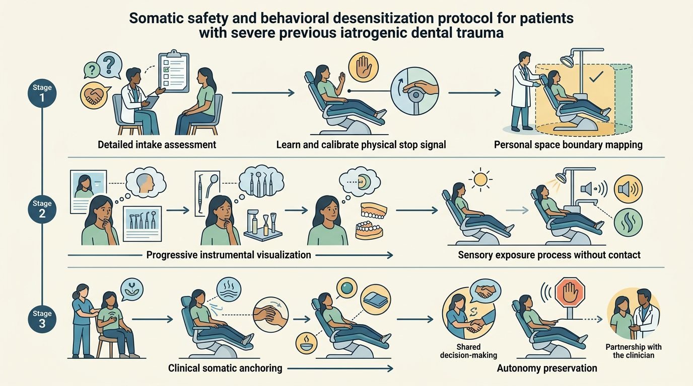
First Visit Protocol: Consultation Outside the Operatory
Rebuilding trust mandates divorcing the initial contact from clinical intervention. The first appointment should take place in a neutral, non-clinical office environment, seated across a conference desk rather than in the dental operatory. White coats should be avoided, and instruments must remain entirely out of sight.
The dialogue centers on taking a detailed trauma history: “Tell me about your past dental experiences, what went wrong, and what specific actions make you feel unsafe.” The practitioner actively listens without interrupting, noting specific sensory triggers like bur sounds, suction tips, or reclining angles. During this visit, no intraoral examination, mirrors, or sharp explorers are introduced unless explicitly requested by the patient. The clinician explains that the patient possesses absolute authority to veto any suggested procedure, establishing that physical force will never be utilized.
Restitution of Autonomy: The Unconditional Stop Signal
Helplessness dissolves only when physical control is returned unconditionally to the patient. Prior to entering the operatory in subsequent appointments, the clinician and patient negotiate an unambiguous non-verbal stop signal, typically raising the non-dominant hand. The protocol is tested repeatedly before any procedure begins.
The dentist instructs: “Raise your hand right now so we can test our response.” When the patient raises their hand, the clinician immediately steps back, lowers their hands, and says, “Whenever that hand moves, everything stops instantly, no questions asked.” Honoring this agreement during clinical treatment is non-negotiable. If a patient signals, the bur or ultrasonic tip must exit the mouth within one second, even if twenty seconds more would complete a cavity preparation. Continuing work after a stop signal instantly reactivates the traumatic memory of entrapment and destroys clinical rapport.
Predictable Infiltration and Objective Anesthesia Verification
Eliminating pain anticipation requires meticulous anesthetic administration combined with objective confirmation of pulpal insensibility before applying mechanical instruments. Topical benzocaine or lidocaine ointment must be applied under dry isolation for two full minutes to anesthetize mucosal nociceptors.
Local anesthetic solutions should be warmed to body temperature (37 degrees Celsius) to reduce injection sting. The clinician delivers the solution using ultra-slow computerized systems or manual micro-injections at rates under one milliliter per minute, mitigating tissue distension pain. Following injection, pulpal anesthesia must be verified objectively using an electric pulp tester or cold thermal spray on the target tooth. Touching the gingiva and asking if it feels numb is inadequate, as soft tissue anesthesia does not guarantee pulpal block. Proceeding to rotate a bur only after the tooth registers zero sensation to freezing stimuli provides physical proof of safety, soothing panic.
Sensory Management and Cognitive Deconditioning
Systematic desensitization requires modifying the sensory environment of the operatory. Ambient noise should be mitigated using active noise-canceling headphones, allowing the patient to listen to podcasts or preferred music. Olfactory triggers like eugenol and acrylic monomers can be masked by diffusing neutral essential oils like lavender in reception areas.
The chair must remain partially upright (at 45 degrees) during conversational check-ins, descending only during operative actions. Overhead lighting should never illuminate the eyes directly; tinted protective eyewear provides physical and psychological shielding. Clinicians employ the “Tell-Show-Do” sequence, explaining each instrument, touching it to the patient’s thumbnail to demonstrate vibration or water spray, and obtaining explicit permission before intraoral entry: “May I place the mirror now?” This choreography restores cognitive control and extinguishes conditioned fight-or-flight reflexes.
Clinical Outcomes and Transformative Practice
Adopting trauma-informed protocols transforms clinical outcomes for chronically phobic populations. Patients who avoided care for decades successfully undergo complex periodontal therapy, crown placements, and endodontic treatments without requiring general anesthesia or deep intravenous sedation.
The investment of additional clinical time during initial visits pays long-term dividends. Chairside emergencies decline, patient loyalty solidifies, and destructive periodontal breakdown is arrested. By understanding the neurobiology of iatrogenic trauma, validating past suffering, and surrendering unilateral procedural control, the dental clinician evolves into a restorative partner, reconstructing oral health and human dignity simultaneously.
References
- Armfield, J. M., & Heaton, L. J. (2013). Management of fear and anxiety in the dental clinic: a review. Australian Dental Journal, 58(4), 390-407.
- Bracha, H. S., et al. (2006). Does “dental phobia” consider posttraumatic stress disorder? Journal of the American Dental Association, 137(12), 1636-1638.
- Hargreaves, K. M., & Keiser, K. (2002). Local anesthetic failure in endodontics: mechanisms and management. Endodontic Topics, 1(1), 26-39.
- Kaysen, D., et al. (2014). Trauma and PTSD: implications for dental care in veteran populations. Journal of Dental Education, 78(8), 1123-1131.
- Raja, S., et al. (2014). Trauma-informed care in dentistry: what, why, and how. Journal of the American Dental Association, 145(11), 1109-1111.
Leave a Reply