
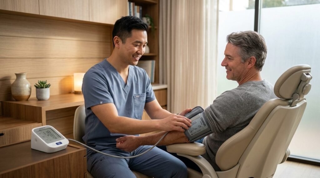
Situational blood pressure elevation triggered by the clinical dental environment, commonly termed white-coat hypertension, represents a frequent clinical dilemma that complicates routine surgical and restorative procedures. When an apprehensive patient enters an operatory, the sight of medical uniforms, sensory operatory odors, or anticipatory fear of needles stimulates acute central autonomic hyperarousal. Peripheral vascular resistance surges, elevating systolic readings above one hundred and forty millimeters of mercury and diastolic pressures above ninety millimeters of mercury in patients who are otherwise normotensive in ambulatory domestic settings. Clinicians often face the difficult dilemma of whether to cancel planned treatments or proceed with local anesthesia containing adrenergic vasoconstrictors. Abruptly dismissing a tense patient reinforces psychological panic and delays essential care, whereas administering unbuffered vasoconstrictors during acute hypertensive spikes risks cardiovascular complications. Resolving this challenge requires dental practitioners to master the neurobiology of situational vasoconstriction, evaluate hemodynamic pharmacological safety objectively, and implement systematic pre-operative calming protocols that restore autonomic equilibrium chairside.
The Autonomic Neurobiology of Situational Vasoconstriction
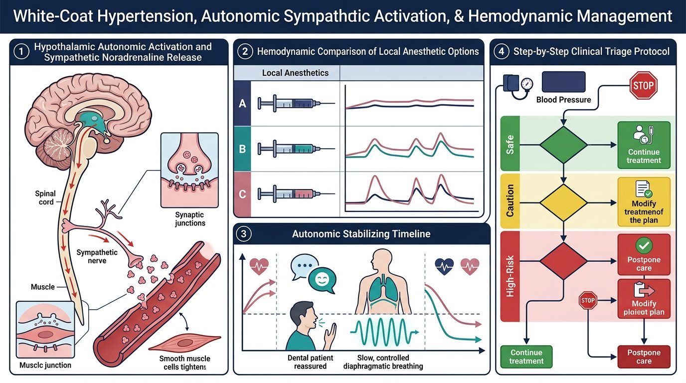
White-coat hypertension represents a hyper-reactive sympathetic defense response mediated by the central autonomic network. Sensory cues associated with the dental clinic activate the basolateral amygdala, which projects dense excitatory efferents to the paraventricular nucleus of the hypothalamus and the rostral ventrolateral medulla. This subcortical activation drives rapid sympathetic discharge through preganglionic splanchnic nerves, stimulating the adrenal medulla to release epinephrine and norepinephrine into systemic circulation.
Circulating catecholamines trigger intense peripheral arteriolar vasoconstriction and a sharp rise in systemic vascular resistance. Concurrently, stimulation of beta-1 adrenergic receptors on myocardial pacemaker cells increases heart rate, while myocardial inotropy accelerates stroke volume. The combined product of elevated cardiac output and constricted peripheral vasculature elevates systemic arterial blood pressure within minutes of the patient crossing the clinic threshold.
Differentiating Situational Reactivity from Sustained Chronic Hypertension
Accurate clinical triage requires distinguishing transient situational spikes from underlying sustained chronic arterial hypertension or hypertensive urgency. Patients with undiagnosed chronic hypertension exhibit persistent structural vascular remodeling, including arteriolar medial hypertrophy, reduced arterial compliance, and end-organ microvascular damage. In contrast, white-coat hypertension is characterized by rapid hemodynamic lability: pressures spike upon operatory entry but drop significantly once the patient feels safe and relaxed.
Relying on a single blood pressure measurement obtained immediately after the patient climbs into the dental chair produces unacceptably high rates of false-positive diagnoses. Physical exertion from climbing clinic stairs, combined with white-coat apprehension, inflates initial readings by twenty to thirty millimeters of mercury. Standard clinical protocols should mandate an initial five to ten-minute stabilization period of quiet, unhurried rest before recording baseline vitals. If initial readings exceed stage two hypertension guidelines, automated oscillometric re-measurement should be repeated after fifteen minutes of calm conversational interaction, allowing sympathetic tone to subside toward baseline levels.
Local Anesthetic Vasoconstrictor Safety: Epinephrine Dynamics
The decision to administer local anesthetic formulations containing vasoconstrictors, such as 1:100,000 or 1:200,000 epinephrine, in patients displaying elevated blood pressure requires precise pharmacological risk assessment. Anxious clinicians often consider utilizing plain mepivacaine or prilocaine without epinephrine, believing that eliminating the pharmacological vasoconstrictor eliminates cardiovascular danger. However, this clinical reasoning contains a fundamental physiological error.
Plain local anesthetics possess intrinsic vasodilatory properties that accelerate intravascular absorption, drastically shortening pulpal anesthesia duration and reducing anesthetic depth. When a procedure begins on an inadequately anesthetized tooth, acute procedural pain stimulates massive endogenous catecholamine release from the patient’s own adrenal medulla. Circulating endogenous epinephrine and norepinephrine levels during acute procedural pain surge to concentrations twenty to forty times higher than the minute amount delivered in a standard dental anesthetic cartridge (0.018 milligrams in 1.8 milliliters of 1:100,000 epinephrine). Providing profound, complete local anesthesia with judicious epinephrine (maintaining the cardiac dose limit of 0.04 milligrams, or two cartridges of 1:100,000) prevents catastrophic endogenous autonomic surges, shielding the cardiovascular system from pain-induced hypertensive crises.
Chairside Somatic Calming and Parasympathetic Reactivation
Down-regulating situational hypertension requires active chairside deployment of somatic calming interventions before picking up a syringe. Because cognitive reassurance alone cannot override limbic sympathetic activation, clinicians must engage physiological feedback loops that stimulate the vagus nerve and activate the parasympathetic branch of the autonomic nervous system. The most effective chairside tool is box breathing or extended-exhalation resonance breathing, in which the patient inhales nasally for four seconds, holds for two seconds, and exhales slowly through pursed lips for six to eight seconds.
Extended exhalation increases intrathoracic pressure, activating aortic and carotid baroreceptors that fire inhibitory signals to the vasomotor center in the brainstem. Parasympathetic efferent impulses travel through the vagus nerve to the sinoatrial node, decreasing resting heart rate and promoting systemic arteriolar vasodilation. Combining structured breathing exercises with dim operatory lighting, calm acoustic environments, and soothing verbal guidance reliably reduces systolic blood pressure by fifteen to twenty-five millimeters of mercury within ten minutes, returning the patient to a medically safe operating window.
Clinical Triage Algorithms and Deferral Thresholds
While humanized calming resolves most instances of white-coat reactivity, clinicians must adhere to clear hemodynamic safety thresholds to protect patient welfare. For elective dental procedures, blood pressure readings should remain below 160/100 millimeters of mercury. If a patient presents with initial readings between 160/100 and 180/110 millimeters of mercury, elective care can proceed safely provided that chairside calming successfully lowers pressures below the 160/100 threshold, treatment is paced conservatively, and anesthetic volume is restricted to two cartridges with vasoconstrictor.
However, if blood pressure readings remain persistently above 180/110 millimeters of mercury despite twenty minutes of quiet rest and calming interventions, elective dental procedures must be deferred. Pressures exceeding this threshold signify severe cardiovascular strain or true hypertensive urgency, where transient surgical stress could precipitate acute myocardial ischemia, cardiac arrhythmia, or cerebrovascular accident. The patient should be referred calmly and promptly to their primary care physician for comprehensive hypertensive evaluation, avoiding alarming language that could exacerbate hypertensive urgency.
Clinical Significance in Humanized Patient Care
Managing white-coat hypertension embodies the integration of hemodynamic science and empathetic clinical presence. Rather than viewing elevated preoperative vitals as a procedural inconvenience or treating the patient as a high-risk obstacle, humanized practitioners recognize situational hypertension as a clear biological signal of acute emotional vulnerability. Combining somatic de-escalation with evidence-based local anesthetic pharmacology guarantees patient safety, fosters profound clinical trust, and ensures safe dental care delivery.
References
- Pickering, T. G., et al. (2002). Ambulatory blood pressure monitoring. New England Journal of Medicine, 354(22), 2368-2374.
- Bader, J. D., et al. (2002). Clinical practice guideline for the management of patients with hypertension in the dental office. Journal of the American Dental Association, 133(12), 172-177.
- Laragh, J. H. (2001). White-coat hypertension: a benign condition or a risk factor? American Journal of Hypertension, 14(9), 839-842.
- Findler, M., et al. (2012). Dental treatment in patients with cardiovascular disease: a guide for dental practitioners. Quintessence International, 43(8), 693-704.
- Perusse, R., et al. (1992). The use of vasoconstrictors in local anesthesia for patients with cardiovascular disease. Fact or fiction? Journal of the American Dental Association, 123(5), 76-82.
Leave a Reply