
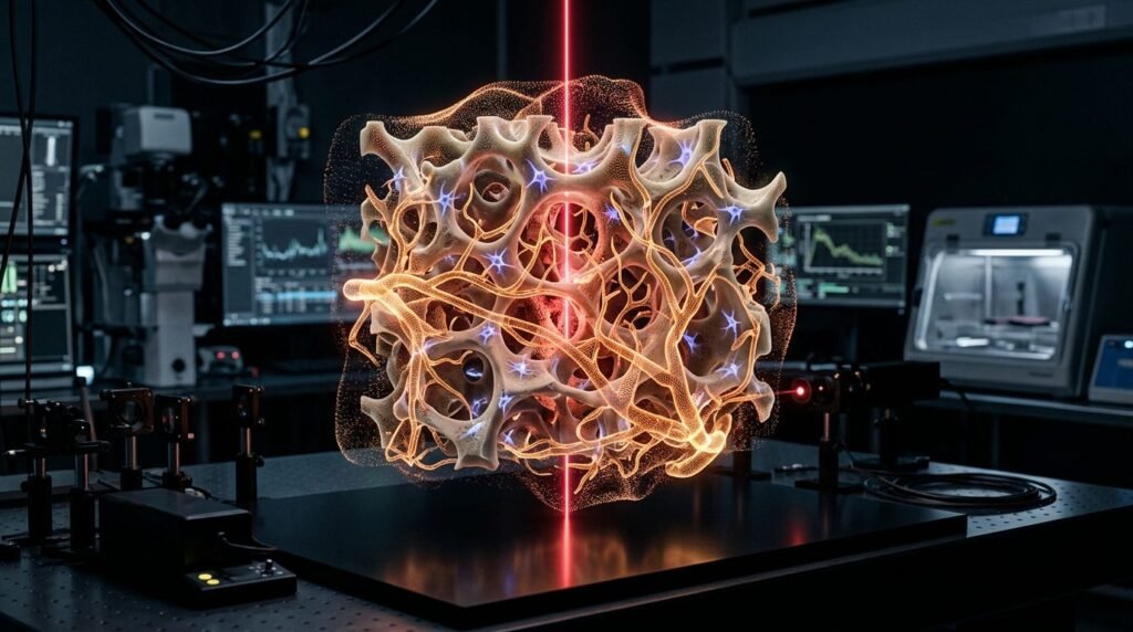
Photobiomodulation acts as an indispensable biophysical adjunct in the management of medication-related osteonecrosis of the jaw by stimulating microvascular angiogenesis, accelerating mucosal epithelialization, and controlling bone marrow inflammation. Medication-related osteonecrosis of the jaw (MRONJ) represents a debilitating complication in patients receiving antiresorptive agents such as zoledronic acid, pamidronate, and denosumab, or antiangiogenic therapies for oncology or osteoporosis. The suppression of bone turnover mediated by osteoclast inhibition, combined with oral microtrauma or tooth extractions, leads to exposed necrotic bone, secondary infection, fistula formation, and refractory pain. Conservative medical therapy frequently fails to achieve complete mucosal closure, whereas extensive surgical resections often induce morbidity in frail patients. The application of low-level laser therapy and surgical erbium laser debridement provides a conservative, tissue-preserving pathway that enhances clinical healing across diverse clinical stages.
Pathophysiological Rationale: Angiogenesis and Cellular Rescue
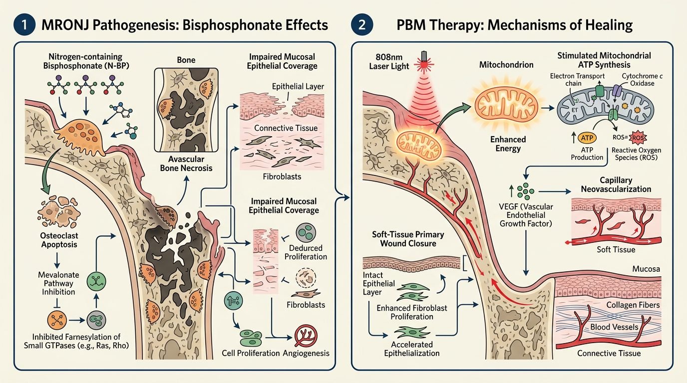
The biological rationale for photobiomodulation (PBM) in osteonecrotic bone lies in overcoming localized tissue ischemia and impaired cellular proliferation. Potent nitrogen-containing bisphosphonates induce apoptosis in osteoclasts, macrophages, endothelial cells, and oral keratinocytes. This cellular toxicity severely impedes the physiological bone remodeling cascade and microvascular perfusion. Irradiation within the red to near-infrared optical window (660 nm to 810 nm) penetrates mucosal and cortical barriers to reach compromised osteocytes and residual mesenchymal stem cells.
At the subcellular level, photons are absorbed by cytochrome c oxidase within the mitochondrial respiratory chain. This photoactivation increases mitochondrial membrane potential, elevates adenosine triphosphate (ATP) synthesis, and generates transient low levels of reactive oxygen species. In turn, these secondary messengers activate transcription factors, including hypoxia-inducible factor 1-alpha (HIF-1alpha) and vascular endothelial growth factor (VEGF). Enhanced expression of VEGF and basic fibroblast growth factor (bFGF) stimulates capillary sprouting at the necrotic periphery. Furthermore, PBM stimulates receptor activator of nuclear factor kappa-B ligand (RANKL) expression in osteoblasts, counterbalancing excessive chemical inhibition and supporting bone turnover.
Diagnostic Staging and Therapeutic Objectives
Clinical staging according to the American Association of Oral and Maxillofacial Surgeons (AAOMS) dictates the therapeutic objective of laser protocols. Stage 0 presents with non-specific bone pain and radiographic sclerosis without clinically exposed bone. In this stage, PBM serves as non-invasive neurovascular therapy, relieving chronic aching and arresting progressive ischemic degradation.
In Stage 1 (exposed, asymptomatic necrotic bone with intact soft tissue borders), PBM aims to induce mucosal overgrowth across minor defects and prevent infectious progression. In Stage 2 (exposed necrotic bone with infection, erythema, and purulent exudate), laser therapy must be combined with targeted systemic antibiotic therapy and antimicrobial photodynamic therapy (aPDT) to resolve localized suppuration before attempting surgical demarcation. Stage 3 (extensive necrosis, pathological fractures, extraoral cutaneous fistulae, or mandibular osteolysis extending to the inferior border) necessitates laser-assisted surgical debridement, where PBM prepares surgical margins and accelerates secondary healing.
Dosimetric Parameters and Clinical Irradiation Protocols
Delivering therapeutic energy into hypovascular bone requires strict adherence to energy density and wavelength selection. Surface biostimulation of intact gingiva and fistula tracts utilizes a dual-wavelength protocol. An initial red diode laser (660 nm, continuous wave, power 50 mW, spot size 0.04 cm2) irradiates the mucosal perimeter to accelerate superficial wound closure. Each point receives an energy density of 4 J/cm2 (fluence) over an exposure duration of 30 to 40 seconds.
To reach deeper cortical compartments and the mandibular canal, clinicians employ an infrared diode laser (808 nm to 830 nm, power 100 mW, spot size 0.2 cm2). The infrared beam is applied perpendicularly around the lesion borders in a contact grid pattern, maintaining a spacing of 5 mm between target points. Each irradiation site receives 6 J/cm2. Total energy per session ranges from 30 to 60 Joules, depending on lesion dimensions. Sessions are repeated twice weekly for six to ten consecutive weeks. Clinicians must avoid excessive thermal accumulation, as power levels exceeding 200 mW or focal overheating induce tissue carbonization and accelerate bone necrosis.
Laser-Assisted Surgical Debridement: The Role of Er:YAG
When necrotic sequestration demands physical removal, the erbium-doped yttrium aluminum garnet (Er:YAG, 2940 nm) laser offers significant clinical advantages over mechanical rotating burs. Mechanical ostectomy using high-speed burs generates microfractures, introduces metal particle contaminants, and produces frictional heat that further compromises marginal vascularity. In contrast, the Er:YAG laser operates via thermo-mechanical micro-explosions within water molecules inside bone hydroxyapatite.
Using a calibrated setting of 200 to 250 mJ per pulse, a frequency of 15 to 20 Hz, short pulse duration (300 microseconds), and continuous sterile saline irrigation (5 to 8 mL/min), the Er:YAG ablation layer-by-layer removes infected and necrotic trabecular bone without thermal alteration. The absence of a thermal coagulative smear layer exposes bleeding bone trabeculae, creating a receptive bed for neo-vascular invasion and platelet clot adherence. Following bone regularization, clinicians apply low-level diode PBM immediately to the surgical cavity prior to tension-free primary mucosal closure.
Antimicrobial Photodynamic Therapy in Refractory Bone Infection
Superficial and deep bacterial colonizations, predominantly involving Actinomyces species, Fusobacterium, and resistant streptococci, exacerbate osteonecrosis by releasing destructive hydrolytic enzymes. Conventional systemic antibiotics often fail to accumulate in therapeutic concentrations within avascular bone sequestra. Antimicrobial photodynamic therapy delivers rapid local bacterial decontamination without selecting antibiotic-resistant clones.
Clinicians infuse a 0.01% methylene blue or toluidine blue O solution directly into bone fissures and fistulous tracts. Following a dark incubation period of three to five minutes to allow photosensitizer uptake across bacterial cell envelopes, the area is irradiated with a red laser (660 nm, continuous wave, 100 mW) for 60 seconds per site. The generated singlet oxygen and hydroxyl radicals destroy bacterial cell membranes, eliminate resistant biofilms, and reduce inflammatory exudate without harming adjacent regenerative osteoblasts.
Clinical Outcomes and Multidisciplinary Practice
Controlled clinical trials and long-term observational cohorts indicate that incorporating photobiomodulation and erbium laser debridement yields complete mucosal healing in over 75% to 85% of stage 1 and 2 MRONJ lesions, substantially surpassing results from antibiotic regimens alone. Furthermore, post-surgical pain scores decrease significantly within the first forty-eight hours, reducing narcotic analgesic reliance.
Success requires close coordination with the patient’s oncologist, endocrinologist, or hematologist. Discontinuing antiresorptive medications before treatment remains a controversial medical decision that must be weighed against skeletal metastatic risk. The dentist provides localized bioenergetic support, protects remaining bone.
References
- Adamo, D., et al. (2020). Low-level laser therapy in the prevention and treatment of medication-related osteonecrosis of the jaw: a systematic review. Supportive Care in Cancer, 28(10), 4589-4601.
- Campisi, G., et al. (2020). Medication-related osteonecrosis of the jaws (MRONJ): an Italian consensus update on behalf of SIPMO and SICMF. Oral Diseases, 26(4), 856-868.
- Ruggiero, S. L., et al. (2022). American Association of Oral and Maxillofacial Surgeons’ position paper on medication-related osteonecrosis of the jaws – 2022 update. Journal of Oral and Maxillofacial Surgery, 80(5), 920-943.
- Vescovi, P., et al. (2014). Surgical approach with Er:YAG laser on bone tissues: our experience in 12 years of clinical practice. Photomedicine and Laser Surgery, 32(9), 503-512.
- Zecha, J. A., et al. (2016). Low-level laser therapy/photobiomodulation in the management of side effects of chemoradiation therapy in head and neck cancer: part 2: proposed applications and specifications. Supportive Care in Cancer, 24(6), 2793-2805.
Leave a Reply